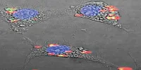
A recent study at the University of Eastern Finland (UEF) has developed a new technique based on bioimpedance that reduces image degradation caused by respiratory motion during a positron emission tomography (PET) scan. The novel technique allows for image reconstruction at a specific phase of the patient's breathing pattern, making it possible to reduce image degradation caused by motion.
A PET scan usually takes several minutes, which is why movement caused by the patient's breathing inevitably degrades image quality. Reduced image quality caused by respiratory motion has been reported to affect PET scanning performed to detect cancer and heart conditions in particular. At worst, image degradation may lead to a wrong diagnosis and inappropriate or unnecessary treatment.
A PET scan usually takes several minutes, which is why movement caused by the patient's breathing inevitably degrades image quality. Reduced image quality caused by respiratory motion has been reported to affect PET scanning performed to detect cancer and heart conditions in particular. At worst, image degradation may lead to a wrong diagnosis and inappropriate or unnecessary treatment.
When synchronising images on the basis of bioimpedance, it was possible to see even smaller details, according to the study presented in the PhD thesis of Tuomas Koivumäki, MSc (Tech.). Motion compensation also significantly influenced the parameters measured from the images. Bioimpedance measurement therefore offers a straightforward technique for acquiring the data needed for motion compensation.
More Accurate Image Acquisition During PET Scans
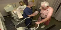
With the newly developed technique, clinicians can have more accurate image acquisition especially during PET scans performed to detect cancers of the chest and upper abdomen, as well as inflammatory diseases of the heart. PET scanning is a modern nuclear medicine imaging method commonly used to detect cancer and heart conditions. Thanks to enhanced image quality, PET images provide new and increasingly accurate data, potentially improving diagnosis reliability and treatment response monitoring.
The newly developed bioimpedance-based technique can be easily integrated into the electrocardiogram (ECG) measurement, which is widely used to monitor heart function during the scan. The UEF study has been published in Medical & Biological Engineering & Computing, Physiological Measurement, and Physics in Medicine and Biology.
Bioimpedance Helps Assess the Patient's Breathing
In techniques based on bioimpedance measurement, a very weak electrical current is passed through the patient's chest, and changes in the resulting voltage are measured. The voltage has been observed to change according to the patient's breathing and cardiac function. Previously, the measurement of bioimpedance has been used as a noninvasive method to assess body composition, blood flow, fluid accumulations in lungs, and indicators of cardiac function.
The UEF study focused on the feasibility of bioimpedance-based measurement techniques for respiratory and cardiac motion compensation in PET imaging. The study first utilised computational models and test subjects to obtain an optimised bioimpedance measurement configuration for simultaneous measurement of respiratory and cardiac gating signals. The second phase of the research focused on analysing whether bioimpedance techniques can be used to reduce respiration-related degradation of PET images.
Source: ScienceDaily.com
Image Credit: Google Images
More Accurate Image Acquisition During PET Scans
With the newly developed technique, clinicians can have more accurate image acquisition especially during PET scans performed to detect cancers of the chest and upper abdomen, as well as inflammatory diseases of the heart. PET scanning is a modern nuclear medicine imaging method commonly used to detect cancer and heart conditions. Thanks to enhanced image quality, PET images provide new and increasingly accurate data, potentially improving diagnosis reliability and treatment response monitoring.
The newly developed bioimpedance-based technique can be easily integrated into the electrocardiogram (ECG) measurement, which is widely used to monitor heart function during the scan. The UEF study has been published in Medical & Biological Engineering & Computing, Physiological Measurement, and Physics in Medicine and Biology.
Bioimpedance Helps Assess the Patient's Breathing
In techniques based on bioimpedance measurement, a very weak electrical current is passed through the patient's chest, and changes in the resulting voltage are measured. The voltage has been observed to change according to the patient's breathing and cardiac function. Previously, the measurement of bioimpedance has been used as a noninvasive method to assess body composition, blood flow, fluid accumulations in lungs, and indicators of cardiac function.
The UEF study focused on the feasibility of bioimpedance-based measurement techniques for respiratory and cardiac motion compensation in PET imaging. The study first utilised computational models and test subjects to obtain an optimised bioimpedance measurement configuration for simultaneous measurement of respiratory and cardiac gating signals. The second phase of the research focused on analysing whether bioimpedance techniques can be used to reduce respiration-related degradation of PET images.
Source: ScienceDaily.com
Image Credit: Google Images
Latest Articles
Cancer, Imaging, PET, scan, bioimpedance
A recent study at the University of Eastern Finland (UEF) has developed a new technique based on bioimpedance that reduces image degradation caused by resp...