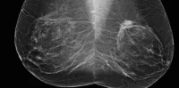
A team of researchers at Rutgers University have developed a new medical imaging method that could help physicians detect cancer and other diseases earlier than before. This could potentially result in speeding up treatment as well as reducing the need for invasive, time-consuming biopsies.
The new imaging method uses nanotechnology to reveal small cancerous tumours and cardiovascular lesions that are deep inside the body. Early tests conducted by Rutgers researchers show that this method has promise.
The fluorescent imaging technique will not only be able to reveal diseases earlier, but will help clinicians learn more about the diseases before they perform surgery. Prabhas Moghe, the lead researcher of this project and a distinguished professor of biomedical engineering and chemical and biochemical engineering, calls it "an optical biopsy".
The technology, developed by Richard Riman, a distinguished professor of Materials Science and Engineering, uses shortwave infrared light. This light penetrates skin and other tissues more deeply as compared to visible light or near-infrared light that are generally used in imaging.
The shortwave infrared light stimulates dyes made with nanocrystals of rare earth elements. To date, fluorescent dyes that react to this light have proven to be too toxic to be used safely and in many cases have been unable to deliver sharp images. However, the dyes that have been developed by Moghe and his team encapsulate rare-earth nanocrystals in a shell of human serum albumin. They are well-tolerated, distribute quickly through the body and accumulate at the disease sites.
Different colours of shortwave infrared light can be used to create a family of probes that would be sensitive to a variety of cancers. This will allow clinicians to get a precise picture of the stage of the disease as well as its makeup.
Initial results of the work have been published in Nature Communications. The Rutgers scientists have also been awarded a $2.2 million grant from the National Institute of Biomedical Imaging and Bioengineering, part of the National Institutes of Health, to further advance their research.
According to Shridar Ganesan, Associate Director for Translational Science at Rutgers Cancer Institute of New Jersey and clinical advisor for the project, "This technique could eventually be used to accurately determine whether a newly detected cancer has spread to nearby lymph nodes, which should help a surgeon deal with the full extent of disease during a single surgery."
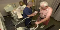
The new method could prove useful since currently surgeons, who are unable to determine how far a cancer has spread, have to do lymph node biopsies. They then have to wait a day for results before they can perform a second surgery. Add the risks, costs and trauma to this mix and it becomes apparent that there is a definite need for a tool that could improve the clinical outcome for cancer patients.
Positive results have already been observed in laboratory mice and the researchers were able to prove that cancer was detected earlier with this method as compared with traditional techniques such as MRI or near-infrared imaging.
Source: Rutgers University
Image Credit: Rutgers University