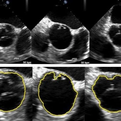
Researchers propose a video segmentation model to recognise regions of interest (ROI) in ultrasounds that are often complicated by low contrast, shadow effects and complex “noise” statistics (unexplained variations).
The research, conducted by a team of primarily mathematicians from Shanghai, Beijing and Singapore, focused their work on the fact that image segmentation, the process of separating a digital image into multiple sections for individual examination, is frequently used in medical image analysis, while real-time applications such as navigation during operational surgery require efficient algorithms.
In their article published in the Society for Industrial and Applied Mathematics (SIAM) Journal on Imaging Sciences, authors Jiulong Liu, Xiaoqun Zhang, Bin Dong, Zuowei Shen and Lixu Gu propose a video segmentation model to recognise ROI in ultrasounds.
“The proposed model aims to track a moving boundary in ultrasound video efficiently and robustly, with a mathematically-sound framework,” said Zhang, an associate professor in the Department of Mathematics and the Institute of Natural Sciences at Shanghai Jiao Tong University.
“Specifically, we tackle the problem by using wavelet frames and incorporating the noise statistics under a variational framework. The continuity and regularity of the moving boundary is effectively incorporated via weighted regularisation, without introducing a heavy computational burden. The overall method can be efficiently solved with a recently-developed fast algorithm, making it useful in real-time clinical applications.”
Multiple published methods of image segmentation currently exist, but Liu et al. specifically implemented variational methods, which are commonly used for motion tracking and edge detection due to their modeling flexibility.
“Variational methods have been demonstrated to be robust and effective for complicated image segmentation tasks,” said Dong, an associate professor of mathematics at Peking University. “The variational framework permits solid theoretical analysis of the models that can well guide the modeling itself and provide fundamental understanding of the solutions.”
Liu et al. also chose to incorporate wavelet frames, which collect more detail than other variational methods and efficiently segment low-quality footage, such as ultrasound video. This is especially true when the image includes features at various scales.
“Wavelet frame regularisation is used because the geometric structures and singularities in different scales can be identified and extracted efficiently from complex noise environments in the wavelet domain,” said Shen, Tan Chin Tuan Centennial Professor in the Department of Mathematics at the National University of Singapore. “It allows us to track and sharpen geometric shapes when they are segmented automatically through sequential images in the video.”
The authors designed their model to segment an ultrasound video both sequentially and collectively. The model incorporates shape priors -- a type of probability distribution -- in single-image segmentation and computes consecutive shape priors automatically for subsequent segmentations.
Liu et al. applied their model to two ultrasound video data sets and obtained numerical results, which confirm the model’s ability to efficiently track ROI. “Ultrasound imaging is an important modality in clinical application due to its low cost and portability,” said Liu, a Ph.D. student at in the Department of Mathematics at Shanghai Jiao Tong University. “However, its related analysis for accurate diagnosis and monitoring is still challenging due to low image quality, artifacts, and noise. The numerical results on real ultrasound data sets demonstrate that the proposed wavelet frame model with distance prior can track the regions of interest effectively, in terms of both segmentation quality and computational time.” The results compare favourably with other approaches.
The model’s success could improve medical approaches and technology that rely on image segmentation, and Liu et al. are looking to expand its use.
“The model can be further extended to other imaging modality or to locate multi-region simultaneously,” added Liu. “More geometric and prior information can be used to enhance the robustness of the method.” Such advancements will continue to increase the speed, efficiency, and performance of image segmentation, the researchers concluded.
Source: Society for Industrial and Applied Mathematics
Journal Reference:
1. Jiulong Liu, Xiaoqun Zhang, Bin Dong, Zuowei Shen, Lixu Gu. A Wavelet Frame Method with Shape Prior for Ultrasound Video Segmentation. SIAM Journal on Imaging Sciences, 2016; 9 (2): 495 DOI: 10.1137/15M1033344


