Executive Summary
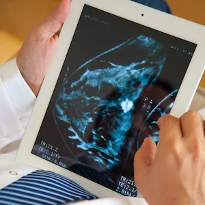
Reading mammograms in a screening setting is one of the most difficult tasks in radiology. Even in a double reader setting where two radiologists rate the same exam, breast cancer is missed relatively often. To improve upon this, computer-aided detection (CAD) systems were developed; however, in practice, the use of CAD marks to highlight suspicious lesions was far from perfect. The large amount of false positive findings marked by the CAD systems were considered to be a distraction and resulted in a perceived low reliability of the systems, and therefore limited use in clinical practice.
You may also like: AI powers radiologists
Understandably, a system that supports radiologists with the decision to refer a woman for further examination would be more effective than a classical CAD-system. Current deep learning systems, for instance, allow determining the probability of a suspicious region to be a carcinoma, whether it is a soft-tissue lesion or calcifications with high accuracy. In addition, several research and commercially available AI-based systems are utilised for mammography analysis. These systems have an accuracy that is on par with that of average, but dedicated, breast radiologists on heterogeneous datasets of mammograms.
However, some radiologists still outperform even these AI-systems. This is likely due to the fact that not all available information is currently being used by these AI-systems. In particular, the temporal information provided by previous studies is not exploited with such systems. By including these factors, the article points out, the performance of AI-systems can be extended beyond the performance of an average breast radiologist.
With the use of MRI, carcinomas can be detected with high sensitivity even for breasts with high density, compared with mammography which is known to be less sensitive for women with high mammography density. The current state-of-the-art breast MRI protocol consists of multiple sequences and lasts about 15 minutes. To make MRI more available for a screening setting, the costs of the technique should be lowered and therefore a lot of research is going into abbreviated MRI-protocols.
Deep learning, according to the article, will contribute significantly to increase the application areas of MRI and make it an economically viable breast cancer screening method. Not only will this open more ways to detect breast cancer earlier and reduce mortality, but will also decrease the variance in performance between radiologists and improve the screening programme as a whole by supporting less experienced radiologists with their decisions.
It should be noted, however, that the applications of AI for breast MRI have not yet left the research domain. Implementing these in clinical practice and proving their efficiency will be a major task for the future, the article concludes.
Source: Full article will be published 22 February 2019 in HealthManagement.org
Image credit: iStock
References:
Latest Articles
MRI, EUSOBI, Radiology, CAD, breast imaging, clinical practice, Radboud UMC, Artificial Intelligence, AI, Clinical Imaging, Mammograms, womens health, deep learning
Executive Summary In this article to appear in HealthManagement the Journal on 22 February 2019, authors Jonas Teuwen, Nikita Moriakov and Ritse Mann pr...


