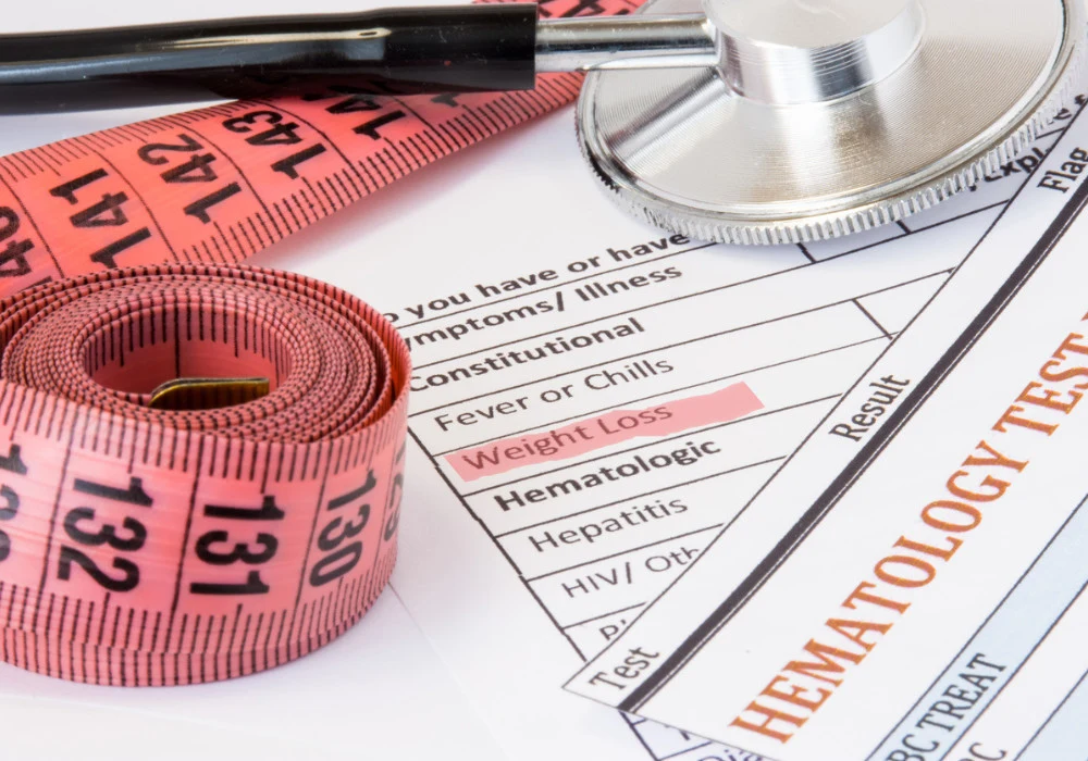
Cancer-associated cachexia (CAC) is a complex syndrome marked by irreversible muscle loss, with or without fat depletion, that cannot be remedied by nutrition alone. Affecting up to 70% of patients with cancer, CAC significantly worsens outcomes by accelerating disease progression, reducing tolerance to treatment and increasing mortality. Despite its prevalence and severity, current diagnostic approaches rely heavily on weight-based metrics, which often fail to capture early-stage changes.
Emerging evidence underscores the need for earlier and more precise diagnostic tools. In this context, medical imaging technologies such as DXA, ultrasound, CT, MRI and PET are being increasingly utilised for their capacity to provide detailed assessments of body composition and metabolic changes associated with CAC. These modalities offer noninvasive, quantifiable insights into muscular and adipose tissue changes, making them instrumental in timely diagnosis and management of the condition.
Dual-energy X-ray Absorptiometry and Ultrasound
Dual-energy X-ray absorptiometry (DXA) is a widely accessible and cost-effective imaging tool that provides detailed measurements of lean mass, fat mass and bone content. It is particularly suited for resource-limited settings due to its short scan times, low radiation exposures and broad clinical availability. DXA is useful in assessing both global and regional muscle and fat distributions, enabling identification of sarcopenia and advanced stages of CAC. However, its diagnostic utility is limited in the early stages of cachexia, as it does not capture metabolic changes or structural alterations within muscle or adipose tissues. Moreover, its spatial resolution and capacity to visualise internal tissue detail are constrained compared to more advanced modalities.
Must Read: Radiology in Early Pancreatic Cancer Detection
Ultrasound (US) imaging offers another accessible, low-cost option that excels in bedside applications. Its portability and absence of ionising radiation make it ideal for critically ill patients. US can detect early muscle changes—such as reductions in muscle thickness, increased echogenicity and decreased elasticity—before overt weight loss becomes apparent. These qualitative and quantitative measurements of muscle properties help to diagnose cachexia in its subclinical phases. However, the utility of US is limited by its dependence on operator skill, lack of standardised imaging protocols and lower reproducibility compared with other modalities. While promising for early detection, further standardisation and training are needed to optimise its role in CAC diagnosis.
CT and MRI in Body Composition and Metabolic Assessment
Computed tomography (CT) offers superior resolution and has become a critical tool in evaluating muscle and adipose tissue alterations in CAC. It allows for precise measurement of skeletal muscle index (SMI) and can detect sarcopenia based on well-established thresholds at specific vertebral levels, such as L3. CT is effective not only in diagnosing muscle depletion but also in assessing fat loss, including in subcutaneous and visceral compartments. Importantly, these tissue reductions can be observed months before CAC is clinically diagnosed. CT also provides insights into muscle quality by quantifying intramuscular fat infiltration, or myosteatosis, which correlates with poorer survival outcomes. Its primary drawbacks include exposure to ionising radiation and high operational costs, which may limit repeat assessments in some clinical contexts.
Magnetic resonance imaging (MRI) complements CT by providing detailed soft tissue contrast without radiation. MRI is capable of detecting early functional and metabolic changes within muscle tissue that precede structural alterations. Techniques such as diffusion-weighted imaging, MR spectroscopy and elastography allow for the assessment of muscle integrity, fat infiltration and tissue stiffness. MRI can also visualise browning of white adipose tissue, a hallmark of CAC-related metabolic reprogramming. However, its clinical use is constrained by high costs, longer scan durations and the need for patient compliance. Ongoing developments in rapid imaging protocols and reconstruction techniques are expected to enhance its utility for early-stage CAC assessment in routine clinical practice.
PET/CT and AI-enhanced Imaging Approaches
Positron emission tomography combined with CT (PET/CT) provides a unique advantage by integrating anatomical and metabolic information. Using tracers like fluorodeoxyglucose (FDG), PET/CT can detect altered glucose metabolism in muscle and adipose tissues, which often precedes weight loss. Elevated FDG uptake in skeletal muscles or adipose depots has been associated with cachexia severity, nutritional status and patient survival. Additionally, PET/CT can monitor brown adipose tissue activity and hepatic metabolic shifts, offering a comprehensive view of systemic changes in CAC. Although PET/CT is a powerful diagnostic tool, its high cost, radiation exposure and limited availability restrict its widespread use.
Recent advancements in artificial intelligence (AI) and radiomics are further enhancing the diagnostic capabilities of imaging in CAC. AI-assisted image analysis can segment muscle and fat compartments with high accuracy and reproducibility, enabling automatic calculation of SMI and fat content. Radiomics can extract complex image features that identify subtle tissue changes indicative of early-stage CAC. Studies have demonstrated the predictive potential of radiomic signatures in anticipating cachexia onset and assessing therapy response. Despite these promising developments, the integration of AI into clinical practice remains limited, necessitating further validation and standardisation before routine adoption.
Early detection and accurate evaluation of cancer-associated cachexia are critical for improving clinical outcomes in patients with cancer. Current diagnostic approaches based on weight and BMI changes are inadequate, particularly in the early stages of the syndrome. Imaging modalities such as DXA, US, CT, MRI and PET/CT offer valuable alternatives by providing comprehensive assessments of muscular and adipose tissue changes, as well as metabolic alterations. Each technique has distinct advantages and limitations, and their combined use can enhance diagnostic accuracy. The integration of AI technologies holds further promise in enabling earlier and more precise detection. As these imaging approaches become more refined and accessible, they are likely to play an increasingly central role in the timely diagnosis and management of CAC.
Source: Radiology: Imaging Cancer
Image Credit: iStock


