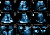
Parathyroid glands are essential regulators of calcium homeostasis, yet their small size and proximity to the thyroid present significant diagnostic challenges. With hyperparathyroidism affecting thousands annually, accurate localisation of parathyroid lesions is critical for guiding effective surgical management. Among various imaging options, ultrasound stands out as a first-line, noninvasive and economical modality for initial evaluation. It enables characterisation of typical parathyroid pathologies and their mimics, often allowing concurrent detection of thyroid abnormalities. Complementary imaging techniques further enhance diagnostic precision, particularly in ectopic or atypical cases.
Ultrasound Applications and Advantages
Ultrasound is highly effective in detecting parathyroid adenomas, the most common cause of primary hyperparathyroidism. Typical adenomas appear as well-defined, hypoechoic lesions with distinctive vascular patterns, such as polar feeding vessels and peripheral arclike flow. The use of high-frequency transducers and Doppler imaging improves sensitivity, especially for lesions under 1 cm. Newer techniques like contrast-enhanced ultrasound and elastography offer additional diagnostic confidence, particularly in distinguishing adenomas from thyroid nodules or lymph nodes.
One of ultrasound’s key advantages is its ability to guide preoperative planning, especially when combined with 99mTc-sestamibi scintigraphy. This combination improves lesion localisation, enabling minimally invasive parathyroidectomy. Additionally, ultrasound facilitates simultaneous evaluation of thyroid nodules, allowing both conditions to be addressed in a single procedure. This shared surgical field between thyroid and parathyroid glands minimises the need for reoperation.
Must Read: AI Copilot for Thyroid Diagnosis: The Impact of ThyGPT
Despite these strengths, ultrasound has limitations. Sensitivity drops for ectopic or intrathyroidal glands and is operator-dependent. Patient anatomy, such as body habitus or kyphosis, can also hinder effective imaging. Therefore, accurate reporting of lesion size, location and characteristics is crucial for informing surgical strategy.
Complementary Imaging Techniques
When ultrasound findings are inconclusive, other modalities help refine diagnosis. 99mTc-sestamibi SPECT/CT remains the most common nuclear medicine approach for parathyroid localisation, particularly effective in identifying ectopic mediastinal glands. Parathyroid tissue retains radiotracer uptake longer than thyroid tissue, aiding differentiation. Combined with ultrasound, this modality offers markedly improved sensitivity.
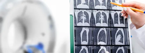
Four-dimensional CT provides a dynamic view of vascular and structural features and is valuable when conventional imaging fails or in cases with prior neck surgery. It is especially useful for detecting ectopic glands and for evaluating complex cases involving distorted anatomy. Dual-energy CT is emerging as a promising technique, using tissue attenuation differences to distinguish parathyroid from thyroid or lymphatic tissue.
Fine-needle aspiration with parathyroid hormone washout can confirm lesion identity when imaging results are ambiguous. Selective venous sampling, while invasive, serves as a last-resort tool in reoperative scenarios or when other localisation attempts fail.
Parathyroid Pathologies and Mimics
Parathyroid adenomas, usually solitary and benign, account for the vast majority of primary hyperparathyroidism cases. Sonographic features often include uniform hypoechogenicity and intense vascularity. Atypical cases may appear cystic, isoechoic or heterogeneous. In some patients, double adenomas or multigland hyperplasia are present, necessitating broader evaluation and surgical consideration.
Parathyroid carcinoma, though rare, tends to present as a larger, irregular and highly vascular lesion with potential for local invasion. Differentiating it from benign adenomas based solely on imaging remains difficult, though features such as capsule irregularity and depth-to-width ratios may provide clues.
Cysts and ectopic glands add complexity to diagnosis. Functional cysts may cause hyperparathyroidism, while ectopic glands may reside in intrathyroidal, mediastinal or carotid sheath locations, challenging ultrasound detection. Additionally, genetic syndromes like multiple endocrine neoplasia may involve all four glands and complicate the clinical picture.
Accurate diagnosis also requires distinguishing parathyroid lesions from mimics. Thyroid nodules, lymph nodes and other structures like the esophagus or longus colli muscle can exhibit overlapping imaging features. Recognising differences in vascular patterns, echogenicity and anatomical continuity helps avoid misdiagnosis. For example, the presence of a polar feeding artery and peripheral arclike vascularity favours a parathyroid adenoma over a thyroid nodule or lymph node.
Ultrasound remains a cornerstone in the assessment of parathyroid disorders, particularly for preoperative localisation of adenomas. When performed by experienced operators, it offers detailed anatomical information, supports surgical planning and facilitates detection of concurrent thyroid disease. Although certain limitations exist—particularly in detecting ectopic or atypical glands—adjunctive imaging methods such as SPECT/CT and 4D CT enhance accuracy. Understanding the characteristic features of parathyroid pathologies and their mimics enables radiologists to deliver precise and effective diagnostic guidance, ensuring optimal outcomes for patients undergoing evaluation for parathyroid disease.
Source: RadioGraphics
Image Credit: iStock


