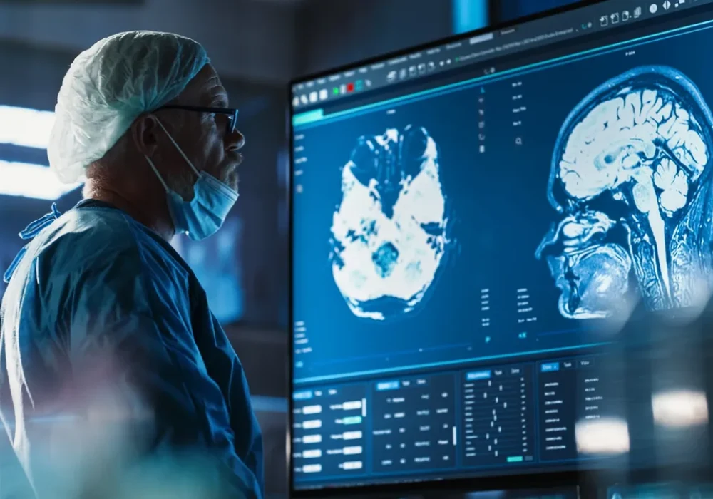
Cancers of the central nervous system (CNS) remain difficult to treat and insufficiently understood, with high morbidity and mortality. Artificial intelligence is moving into the neuro-oncological pathway, spanning prevention, diagnosis, treatment, prognostication and rehabilitation. Reported uses include image analysis, molecular characterisation, biomarker discovery, target identification, tailored management and neurorehabilitation. Alongside this progress are persistent gaps in data access and quality, limited prospective validation, operational barriers to implementation and regulatory and ethical considerations. Near-term gains are most likely where validated AI tools are scaled for specific tasks, while broader approaches may mature as technical, data and governance challenges are addressed.
Imaging and Diagnostic Precision
Clinical magnetic resonance imaging (MRI) underpins detection, characterisation and planning. AI trained on structural MRI has been applied to denoising, registration, artefact correction, detection, segmentation, classification and grading. Multicentre evaluations indicate benefits for classification accuracy and volumetric assessment, though many results come from relatively small or homogeneous cohorts. Routine adoption will depend on prospective, continuous, multicentre validation and harmonised acquisition and processing.
Non-invasive inference of molecular features that influence management has attracted interest. Radiomics, radiogenomics and deep learning using MRI and positron emission tomography (PET) have reported promising performance, but tissue-based corroboration and biological annotation remain essential to ensure clinical utility.
Diagnostic scope is also expanding with modalities not normally interpreted by humans. Examples include ultrasound radio frequency signals for intraoperative molecular diagnosis and functional MRI biomarkers linked to glioma grade. Generative models can synthesise images when data are scarce or noisy, such as super-resolved spectroscopy or de novo post-contrast MRI from pre-contrast scans, although association-based methods have inherent limits without mechanistic grounding. Physics-constrained or biophysical modelling offers an alternative path to realistic synthetic data and explainability, including in chemical exchange saturation transfer MRI.
Digital neuropathology shows similar momentum and obstacles. Deep learning on whole-slide images supports localisation, segmentation, grading and molecular prediction and rapid intraoperative classification using label-free optical techniques or methylation profiles can aid margin assessment and subtype identification. However, digitisation and labelling are resource intensive, datasets vary across institutions and inter-rater variability persists. Foundation models trained in a self-supervised manner may scale autolabelling and improve generalisability, yet resource needs and regulatory and ethical issues limit near-term deployment.
Treatment Planning and Target Discovery
In radiotherapy, AI is being used for automatic segmentation of organs at risk to improve efficiency and consistency. Work is extending to tumour target delineation, including gliomas and brain metastases. The clearest gains appear in anatomy without major distortion, while infiltrative tumours highlight the need for standardised algorithms and imaging inputs. Intraoperative support is also advancing, with AI-assisted feedback from operative video and imaging to highlight regions more likely to contain tumour, interpret signals beyond the immediate field and flag unnecessary risks. Rapid intraoperative molecular diagnosis based on optical imaging or methylation data can further guide resection decisions. Robotic systems for stereotactic sampling and image-guided microsurgery have shown non-inferiority to conventional techniques, indicating potential to reduce human error in high-stress environments.
Systemic therapy for aggressive entities such as glioblastoma and diffuse midline glioma remains constrained by limited responsiveness to chemotherapy and the absence of recently approved agents. AI-driven stratification aims to identify patient subsets more likely to benefit from specific interventions. Stemness predictors derived from transcriptomic profiles have been associated with susceptibility to programmed death-ligand 1 inhibition, differences in overall survival and distinct immune microenvironments. Proteomic axes linked to aggressiveness and chemoresistance implicate alternative immunological targets beyond established markers. Network-based kinase–substrate analyses highlight master kinases in functional niches, suggesting candidates for targeted intervention. Coupling such target discovery with AI-driven de novo compound design could accelerate development, although clinical effectiveness has yet to be demonstrated.
Must Read: Enhancing Oncology with AI: UCSF’s Scalable Support System
Risk Stratification, Response Assessment and Rehabilitation
AI-supported risk models integrate histomolecular heterogeneity, imaging features and cytoarchitectonic patterns to complement established prognostic factors. Translational inference from MRI and diffusion imaging has been associated with predictions of overall and progression-free survival, and image-derived risk scores correlate with differentially enriched pathways and signatures. Refinements within tumour classes underscore the need for more granular stratification: epigenetic subclasses in high-grade astrocytoma with piloid features and diffuse midline glioma show divergent outcomes and biology, while in glioblastoma a high-neural subgroup characterised by synaptic integration correlates with worse survival and less benefit from near-complete resection. Mapping regional gene expression programmes to morphology, connectivity and signalling is opening routes to spatially resolved biomarker discovery across magnetoencephalography, digital pathology and serum.
Reliable response assessment is central to decision-making. AI-based volumetric quantification reduces inter-rater variability in estimating time to progression and improves consistency over conventional criteria. Integrated analyses combining contrast-enhanced MRI with radiotherapy dose distributions show potential to predict local control after stereotactic radiosurgery. Distinguishing pseudoprogression from true progression can be aided by AI to avoid premature changes in therapy. Response-adaptive radiotherapy is under exploration, with iterative imaging informing adjustments during treatment, subject to stronger evidence on biological relevance. Management of late toxicities, including cognitive dysfunction and radiation necrosis, may benefit from AI-assisted planning to spare organs at risk, non-invasive monitoring and prediction of radiotoxicity-associated gene expression patterns.
Neurorehabilitation presents further opportunity. AI-enabled brain–computer interfaces, both invasive and non-invasive, have demonstrated decoding of intended movements and translation of neural activity into speech, indicating potential to mitigate tumour- and treatment-related neurocognitive and motor impairments. Generative models for neural stimulation or interface control could support restoration of complex functions in long-term survivors with persistent deficits.
AI is reshaping neuro-oncology across non-invasive precision diagnosis, digital pathology, intraoperative guidance, radiotherapy planning, target discovery, prognostication, response assessment and rehabilitation. Impact will depend on improved data collection and annotation, harmonised pipelines and prospective multicentre validation linked to meaningful outcomes. Regulatory and ethical frameworks need to keep pace with generalist models and synthetic data while enabling safe deployment. Progress is most likely where validated tools for defined tasks are scaled, with careful exploration of foundation models, biophysical approaches and integrative stratification aligned to patient benefit.
Source: The Lancet Digital Health
Image Credit: iStock


