


An imaging study in Canada has found that MRI and CT could help explain the symptoms and exercise capabilities of people with mild-to-moderate chronic obstructive pulmonary disease (COPD). Researchers said the findings, published in the journal Radiology, point the way to better treatment for some COPD patients.
“COPD is a very heterogeneous disease,” said study co-author Grace Parraga, PhD, from the Robarts Research Institute in London, Ontario, Canada. “Patients are classified based on spirometry, but patients with the same air flow may have different symptoms and significant variation in how much regular activity they can perform, such as walking to their car or up the stairs in their home.”
Spirometry is a lung function test — in which the patient takes a breath and exhales forcefully into a tube connected to a machine — that is commonly used to help diagnose COPD. This test produces a figure called the forced expiratory volume in one second (FEV1). According to researchers, FEV1 does not necessarily reflect the whole picture of the lungs, but airways disease and emphysema can be directly measured with imaging.
For the study, Dr. Parraga and colleagues performed conventional CT and inhaled noble gas MRI (a technique for visualising air spaces in the lungs) on 116 people with COPD, including 80 with milder disease. The patients also underwent lung capacity testing, filled out a quality of life questionnaire and took a six-minute walk to measure their exercise tolerance over a short period of time.
The researchers found that in mild-to-moderate COPD patients with modestly abnormal FEV1, MRI measurements of emphysema were strongly correlated with exercise limitation, while both CT and MRI measurements of emphysema helped explain symptoms.
The implications of the findings are significant for patients with mild COPD and abnormal FEV1, according to Dr. Parraga. “FEV1 doesn’t tell the whole story,” she noted. “With lung imaging, we can look at patients with mild disease much more carefully and change treatment if necessary.”
Findings associated with COPD include emphysema, or damage to the air sacs in the lungs that prevents people from getting the oxygen they need, especially during exercise. While no cure exists for emphysema, there are steps people can take to mitigate symptoms. Emphysema is under-recognised as a source of COPD, Dr. Parraga said, and as a result, patients may be getting suboptimal treatment.
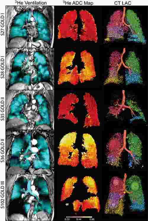
Figure 1. Images in representative patients with mild-to-moderate or severe COPD.
Source and image credit: RSNA
“COPD is a very heterogeneous disease,” said study co-author Grace Parraga, PhD, from the Robarts Research Institute in London, Ontario, Canada. “Patients are classified based on spirometry, but patients with the same air flow may have different symptoms and significant variation in how much regular activity they can perform, such as walking to their car or up the stairs in their home.”
Spirometry is a lung function test — in which the patient takes a breath and exhales forcefully into a tube connected to a machine — that is commonly used to help diagnose COPD. This test produces a figure called the forced expiratory volume in one second (FEV1). According to researchers, FEV1 does not necessarily reflect the whole picture of the lungs, but airways disease and emphysema can be directly measured with imaging.
For the study, Dr. Parraga and colleagues performed conventional CT and inhaled noble gas MRI (a technique for visualising air spaces in the lungs) on 116 people with COPD, including 80 with milder disease. The patients also underwent lung capacity testing, filled out a quality of life questionnaire and took a six-minute walk to measure their exercise tolerance over a short period of time.
The researchers found that in mild-to-moderate COPD patients with modestly abnormal FEV1, MRI measurements of emphysema were strongly correlated with exercise limitation, while both CT and MRI measurements of emphysema helped explain symptoms.
The implications of the findings are significant for patients with mild COPD and abnormal FEV1, according to Dr. Parraga. “FEV1 doesn’t tell the whole story,” she noted. “With lung imaging, we can look at patients with mild disease much more carefully and change treatment if necessary.”
Findings associated with COPD include emphysema, or damage to the air sacs in the lungs that prevents people from getting the oxygen they need, especially during exercise. While no cure exists for emphysema, there are steps people can take to mitigate symptoms. Emphysema is under-recognised as a source of COPD, Dr. Parraga said, and as a result, patients may be getting suboptimal treatment.
Figure 1. Images in representative patients with mild-to-moderate or severe COPD.
Source and image credit: RSNA
References:
Parraga
G, et al. (2015) COPD: Do Imaging Measurements of Emphysema and Airways Disease Explain Symptoms and Exercise Capacity? Radiology, July 2015.
Latest Articles
healthmanagement, COPD, imaging, spirometry, MRI, CT, lung
An imaging study in Canada has found that MRI and CT could help explain the symptoms and exercise capabilities of people with mild-to-moderate chronic obstructive pulmonary disease (COPD).
, professor of radiology and surgery at Dartmouth")


