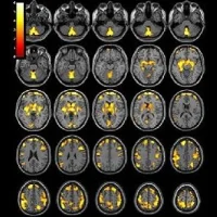
Scientists are developing a new molecular, preclinical hybrid imaging system that combines positron emission tomography (PET) and MRI, and can help experts to find new drugs and advance imaging research. The new scanner includes a window for tissue observation, and an imaging chamber.
The hybrid scanner, presented at the 2015 Annual Meeting of the Society of Nuclear Medicine and Molecular Imaging (SNMMI), is intended for research into the microenvironment of tumours and other tissues, and co-registration of multiple lines of imaging data, according to researchers.
The system combines PET for physiological information from radiotracers, conventional and hyperpolarised magnetic resonance imaging for soft-tissue contrast and tracking of minute biochemistry. It also includes luminescence, fluorescence and optical imaging for investigations by microscope.
The researchers presented a study in which a tumour cell line was transplanted into a rat. The animal was then imaged with conventional MRI, hyperpolarised MRI, a positron detector, and a luminescence sensor, and tissues analysed using a fluorescence microscope.
"This technology allows us to obtain in-depth knowledge of molecular imaging techniques, how to optimise them, and how to leverage data with statistical analysis while advancing new radiotracers and contrast agents for the imaging and treatment of a range of diseases," said the study's lead author Zhen Liu, PhD candidate, nuclear medicine department, Technical University Munich (Germany).
Understanding the physiology behind multimodal imaging can be difficult given discrepancies between macroscopic and microscopic images and between images of extracted or transplanted tissues versus images of a live subject, Liu explained. The development of high-resolution multimodal intra-vital imaging "can bridge these discrepancies and offer a tool for the long-term observation of underlying physiology,” Liu added.
SNMMI is an international scientific and medical organisation dedicated to raising public awareness about nuclear medicine and molecular imaging, a vital element in today’s medical practice that changes the way common and devastating diseases are understood and treated. The society's annual meeting is a highly anticipated event as this showcases new research and technology.
Source and image credit: Society of Nuclear Medicine and Molecular Imaging
Latest Articles
healthmanagement, MRI, PET, SNMMI, hybrid scanner, molecular imaging
Scientists are developing a new molecular, preclinical hybrid imaging system that combines positron emission tomography (PET) and MRI, and can help experts to find new drugs and advance imaging research.


