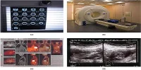
According to a new study published in the Journal of Neurotrauma, Diffusion Tensor Imaging (DTI), a specialised magnetic resonance imaging (MRI) technique that detects microstructural changes in brain tissue, can help physicians better predict the likelihood of poor clinical outcomes following mild traumatic brain injury as compared to other conventional imaging techniques such as a CT scan.
During the study, DTI
for white matter injury was used in 76 adult mild traumatic brain injury
patients at the semi-acute stage (11.2±3.3 days). Both the whole-brain
voxel-wise and region-of-interest (ROI) approaches were used. In order to
evaluate the clinical relevance of DTI, the study team evaluated correlations
between three- and six-month outcomes and imaging, demographic/socioeconomic, and
clinical predictors. In the study, DTI demonstrated utility in an inclusive
group of patients with heterogeneous backgrounds and also in patients without
substance abuse or neuropsychiatric history.
An estimated 75 percent of the
1.7 million patients in the US seek medical attention for mild traumatic brain
injury (mTBI). mTBI refers to a non-penetrating head trauma that results in
confusion, disorientation, loss of consciousness, post-traumatic amnesia and
transient focal neurological signs or seizure. In order to provide optimal care
to patients who experience an acute head injury, it is important to predict
which of these patients are likely to suffer ongoing dysfunction three to six
months down the road.
The results from this
study have been presented by Esther Yuh and co-authors from the University of
California, San Francisco, Erasmus MC-University Medical Centre (Rotterdam, The
Netherlands), Mount Sinai School of Medicine (New York, NY), Seton Brain and
Spine Institute (Austin, TX), University of Pittsburgh Medical Centre (PA),
University of Texas (Austin), Antwerp University Hospital (Edegem, Belgium),
and University of Cambridge Addenbrooke's Hospital (Cambridge, UK). It is the
first published study that compares DTI to conventional imaging. The findings
show that there were significant differences between the white matter of mTBI
patients who had positive versus negative findings on the CT scan and MRI
evaluation.
According to John T.
Povlishock, PhD, Professor, Medical College of Virginia Campus of Virginia
Commonwealth University, Richmond and the Editor-in-Chief of the Journal of
Neurotrauma, "This exceptionally well done study addresses an issue of
continuing controversy and confusion. The authors make an extremely important
observation that MRI studies, including DTI parameters, are integral in
informing prognosis after mild TBI. When taken together with the other
publications from the TRACK-TBI Study Group, these findings should prove
invaluable in assessing the occurrence of mild TBI and informing patient
outcome."
Source: Eurekalert!
Image Credit: Wikimedia Commons