![Tuberculosis Diagnostics: The Promise of [18F]FDT PET Imaging](https://res.cloudinary.com/healthmanagement-org/image/upload/c_thumb,f_auto,fl_lossy,h_184,q_90,w_500/v1721132076/cw/00127782_cw_image_wi_88cc5f34b1423cec414436d2748b40ce.webp "Tuberculosis Diagnostics: The Promise of [18F]FDT PET Imaging")


Currently being developed by University of Washington scientists and engineers, a novel, credit-card sized, low cost device could assist pathologists in their analysis of biopsies and the subsequent diagnosis of pancreatic cancer in an earlier and faster manner than ever before.
The prototype is able to conduct basic steps required for processing a biopsy by relying fluid transport instead of human hands to deal with the tissue.
Of all cancers, pancreatic cancer is an exceptionally devastating disease. At least 94% of patients will succumb within five years, and it was ranked in the top ten deadliest cancers in 2013.
While routine screenings for breast, colon and lung cancers have improved treatment and outcomes for patients with these diseases due to early detection, many behavioural facts of pancreatic cancer are unknown. This leads to patients often receiving a diagnosis when the disease has already progressed too far.
The research team presented its initial results at this month’s SPIE Photonics West conference and recently filed a patent for this first-generation device and future technology advancements.
Eric Seibel, a UW research professor of mechanical engineering and director of the department's Human Photonics Laboratory is confident that this new process will assist pathologists in making a more rapid diagnosis and allow for to improved prognosis as they will determine more accurately how invasive the cancer has become.
In essence, the innovative device would turn the pathology lab’s manual and time-consuming process into an automatic and streamlined process. At present, a pathologist takes a biopsy tissue sample which is sent to the lab where it will be cut into thin slices, stained and put on slides in order to be analysed for abnormalities optically in 2-D to diagnose cancer.
As explained by Ronnie Das, a UW postdoctoral researcher in bioengineering who is the lead author on a related paper, the UW's technology would process and analyse whole tissue biopsies for 3-D imaging, offering a more complete picture of the cellular makeup of a tumor. When a piece of tissue is cut, information about it is lost. By keeping the original tissue biopsy intact, the abnormal cell growth can be viewed in its entirety, and connections, cell morphology and structure as it looks in the body, can be seen.
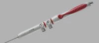
The team is building a thick, credit card-sized, flexible device out of silicon that allows a piece of tissue to pass through tiny channels and undergo a series of steps that replicate what happens on a much larger scale in a pathology lab. The invention harnesses the properties of microfluidics, enabling tissue to move and stop easily through small channels without the application of a lot of external force. Furthermore, it avoids clinicians handling the tissue; instead, a tissue biopsy taken with a syringe needle could be deposited directly into the device to begin processing.
According to the researchers this is the first time material larger than a single-celled organism has successfully moved in a microfluidic device. This achievement could have implications across the sciences by automating analyses that are customarily done by humans.
Das and Chris Burfeind, a University of Washington undergraduate student in mechanical engineering, designed the device by building a mold using a petri dish and Teflon tubes. The following step was to pour a viscous, silicon material into the mold, creating a small, transparent instrument with seamless channels that are both curved and straight.
The next objective is to combine all of the steps into a more robust device to include 3-D imaging, and build and optimise it for use in a lab. Future developments could include layers of channels that would allow more analyses on a piece of tissue without adding more bulk to the device.
The UW researchers say the technology could be used as an over-the-counter kit to process biopsies, then that information could be sent to pathologists who could examine it for signs of cancer from remote locations. Additionally, Das said it could potentially reduce the time it takes to diagnose cancer to a matter of minutes.
The image shows this prototype of a microfluidic device that has both curved and straight channels for transporting tissue biopsies. The silicon material is lightweight, flexible and transparent.
Source and photo: Science Daily
7 February 2014
Latest Articles
Cancer, Research, Medical Devices, 3D imaging, tissue, Biopsy, cancer prevention, cancer diagnosis
Currently being developed by University of Washington scientists and engineers, a novel, credit-card sized, low cost device could assist pathologists in th...