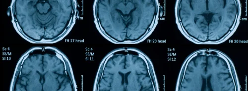
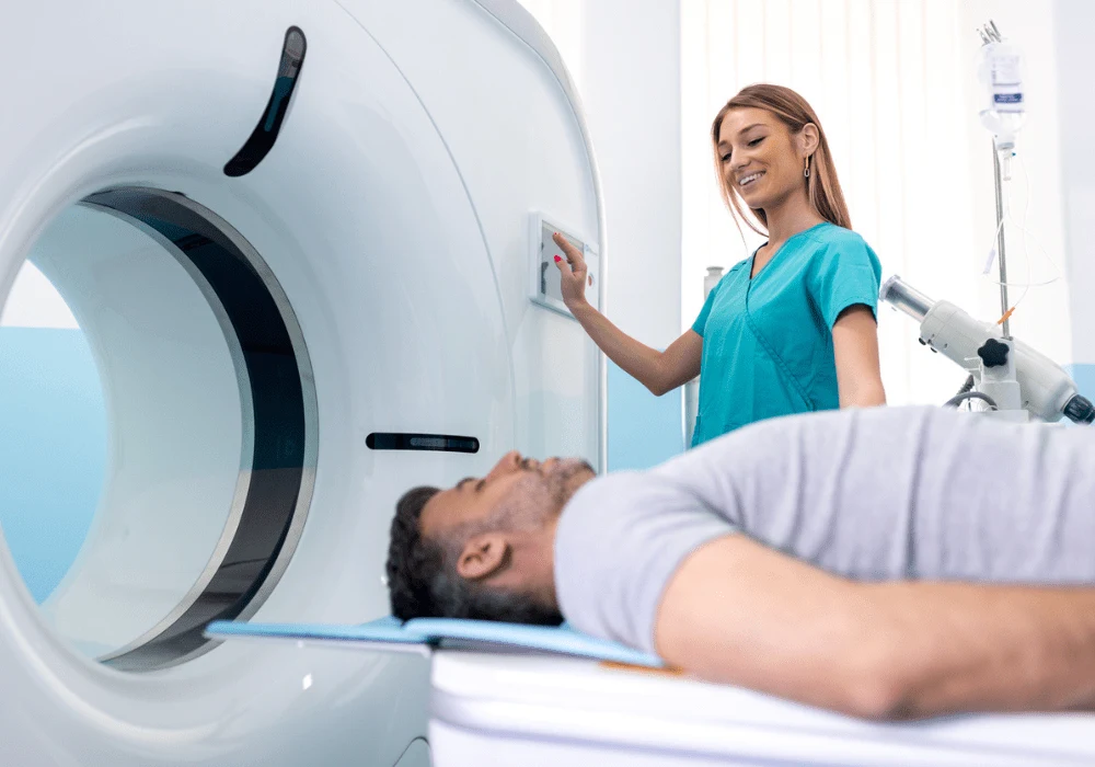
Magnetic Resonance Imaging (MRI) is a cornerstone of modern medical diagnostics due to its non-invasive nature and lack of ionizing radiation. While it is generally considered a safe procedure, several risks are associated with the powerful electromagnetic fields generated during an MRI scan. These fields, essential for creating detailed images, pose potential hazards, including projectile risks, heating, and tissue damage. Understanding and mitigating these risks is critical for ensuring the safety of both patients and healthcare professionals. This ESR Essentials practice recommendation published in European Radiology outlines essential MRI safety principles, focusing on the nature of these risks and recommendations for managing them effectively.
The Static Magnetic Field (B0) and Its Hazards
The MRI environment is dominated by a strong static magnetic field, often called B0. This field, always present in MRI scanners, is typically 1.5 to 3 Tesla (T) in clinical settings but can reach up to 8 T in research environments. The static field’s strength is crucial for image quality but also presents significant safety concerns. One of the primary risks is the projectile hazard, where ferromagnetic objects are drawn toward the scanner with great force, potentially causing injury or damage. These objects can become projectiles, especially near the scanner’s bore, where the magnetic field is strongest. It is imperative that only authorized personnel, well-trained in MRI safety, have access to the MRI room to prevent such accidents.
Beyond external objects, internal ferromagnetic materials, such as surgical implants or shrapnel, pose risks. The spatial field gradient, which measures the change in magnetic field strength across space, can cause ferromagnetic implants to move or twist within the body. This could lead to tissue damage, especially if the object is near sensitive areas like nerves or blood vessels. To mitigate these risks, strict adherence to safety protocols is essential, including careful screening for implants and using equipment labelled as MRI-safe or MRI-conditional.
Radiofrequency (RF) Fields and Tissue Heating
During an MRI scan, radiofrequency (RF) fields, represented as B1, are used to excite hydrogen nuclei in the body, generating the images. However, these fields also transfer energy to the body, resulting in tissue heating. The amount of heating is measured by the Specific Absorption Rate (SAR), typically set at 2 W/kg or 4 W/kg, depending on the scanning mode. While SAR limits are designed to protect patients, certain conditions can increase the risk of burns or other injuries.
For example, conductive materials like electrodes, damp clothing, or sweat can form electrical loops, leading to localised heating and burns. Implants, particularly those with long, conductive leads, can also resonate with RF fields, further increasing the risk of tissue heating. The resonance length of these implants depends on the surrounding tissue and the MRI’s frequency, making it difficult to predict when heating will occur. Additionally, proximity burns can occur when the patient’s body touches the scanner bore. To prevent these injuries, padding must be used to ensure no direct contact between the patient and the scanner.
RF-induced heating is particularly problematic for patients with tattoos or metallic implants, as these can act as antennas, exacerbating the heating effect. A thorough risk assessment, including the implant’s material and positioning considerations, is critical to avoid such complications. Adhering to manufacturer guidelines on implant safety and scanning modes can help mitigate these risks, ensuring a safe MRI experience for patients with implants.
Time-Varying Gradient Fields and Associated Risks
In addition to the static and RF fields, MRI scanners use time-varying gradient fields (dB/dt) to spatially encode the MR signal. These gradients, essential for producing detailed images, generate loud noises during the scan, often reaching levels that require hearing protection. Patients and staff in the MRI room must use earplugs or earmuffs to avoid potential hearing damage.
The time-varying gradients can also induce peripheral nerve stimulation (PNS), which patients experience as a tingling sensation. While PNS is generally harmless, it can be uncomfortable and, in rare cases, may interfere with patient cooperation during a scan. Modern MRI machines offer noise-reduction technologies and operate in different modes to manage the risk of PNS, but it remains an essential consideration during the scan planning process.
For patients with implants, the time-varying fields present additional concerns. These gradients can induce electrical currents in conductive implants, potentially leading to tissue heating or interference with the device’s function. Devices like pacemakers, neurostimulators, and other active implants are particularly vulnerable to these effects. It is essential to evaluate the compatibility of any implanted devices with the MRI’s operating conditions before conducting the scan. This includes checking for specific safety guidelines provided by the device manufacturers, such as acceptable dB/dt limits.
Conclusion
MRI is a powerful diagnostic tool, but it is not without risks. The static magnetic field, radiofrequency field, and time-varying gradient fields used in MRI scanning all pose potential hazards, including projectile risks, tissue heating, and peripheral nerve stimulation. These risks, while significant, can be effectively managed through thorough screening, adherence to safety protocols, and proper training of all MRI personnel.
Healthcare professionals must ensure that patients are carefully screened for contraindications, such as the presence of ferromagnetic objects or implants, before undergoing an MRI scan. Risk assessments should be performed and documented, weighing the benefits of the scan against the potential risks. For patients with implants, special care should be taken to adhere to manufacturer guidelines and ensure that the MRI scanner’s operating conditions are compatible with the device.
In conclusion, while MRI remains one of the safest imaging modalities available, maintaining a high standard of safety requires constant vigilance, strict protocols, and ongoing education. By doing so, healthcare providers can ensure that MRI continues to provide invaluable diagnostic information with minimal risk to patients and staff.
Source Credit: European Radiology
Image Credit: iStock


