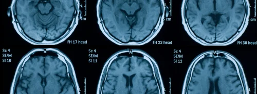
Magnetic Resonance Imaging (MRI) is a vital diagnostic tool that provides detailed images of internal structures, especially the brain. However, traditional MRI processes have limitations, such as long scan times and image noise, which can degrade image quality. Parallel imaging techniques have helped accelerate MRI scans, but at high acceleration rates, the images often suffer from noise and artefacts. To overcome these issues, new artificial intelligence (AI) advancements have emerged, offering solutions that can significantly enhance MRI imaging. A recent article published in Radiology Advances explores how a deep-learning denoising technique can improve the quality of highly accelerated parallel MRI scans, focusing on brain imaging while reducing scan times without sacrificing diagnostic accuracy.
AI-Based Denoising in MRI: An Overview
Parallel imaging techniques, such as Sensitivity Encoding (SENSE) and Generalised Autocalibrating Partially Parallel Acquisitions (GRAPPA), have long been used to accelerate MRI by acquiring multiple data channels simultaneously. However, these methods introduce noise and aliasing artefacts as acceleration factors increase. Deep learning has emerged as a solution to this problem, specifically in denoising noisy images. Unlike conventional methods that rely on proprietary raw data for image reconstruction, AI-based denoising techniques operate within the image domain. This approach simplifies the process and makes it applicable across multiple MRI vendors.
The AI-based denoising model described in this study operates as a two-stage process. In the first stage, MRI images are acquired using a high acceleration factor. Due to undersampling, these images often suffer from noise and minor artefacts. The second stage applies a deep-learning model to correct these distortions. By training on a wide variety of images with simulated noise patterns, the model can eliminate noise while preserving essential anatomical details, resulting in images of high diagnostic quality.
Clinical Evaluation and Data Processing
The evaluation of this AI-based denoising model was carried out using both retrospective and prospective data. Retrospective data, covering a six-year period, were sourced from over 2000 MRI studies, while prospective data were collected from 200 subjects in a clinical setting. These datasets included images from various scanner models and vendors, ensuring the generalizability of the results. The AI model was trained to handle common types of degradation, such as Rician noise and aliasing artefacts, which often accompany accelerated parallel imaging.
To ensure the validity of the model's performance, it was tested on a subset of prospective scans, comparing high-acceleration, AI-enhanced images with baseline low-acceleration images. The clinical evaluation focused on metrics like signal-to-noise ratio (SNR), contrast-to-noise ratio (CNR), and spatial resolution. Five radiologists reviewed the enhanced images, assessing both qualitative and quantitative metrics to confirm the efficacy of the AI model in reducing noise and improving image quality.
Key Results and Findings
The study's results demonstrated significant improvements in both image quality and efficiency. On average, the scan time for the accelerated sequences was reduced by approximately 29%, with scan time reductions ranging from 19% to 41%, depending on the sequence and scanner model. The AI-enhanced images consistently improved SNR, CNR, and spatial resolution despite the higher acceleration factors. The AI-enhanced images often surpassed the quality of standard low-acceleration images in terms of diagnostic clarity.
One of the most compelling findings was the ability of the AI-enhanced images to retain, or even improve, the visibility of critical anatomical structures and pathologies. Radiologists rated the AI-enhanced images as non-inferior or superior to the baseline images for the majority of qualitative criteria, including image noise, artefact reduction, and pathology visibility. The AI model was particularly effective at preserving the clarity of small anatomical structures, such as sulci and white matter lesions, which are crucial for accurate diagnosis in brain imaging.
Quantitative measures of SNR and CNR further supported these findings. The AI-enhanced images showed a significant improvement in SNR, increasing from an average of 39 in the input images to 64 in the enhanced images. Similarly, CNR values rose from 11 to 16 after AI processing. These improvements are critical for maintaining the diagnostic integrity of the images, as higher SNR and CNR values translate into clearer and more distinguishable anatomical features.
Challenges and Future Directions
While the study's results were promising, there are still challenges and limitations to address in the future. The AI-based denoising model was only tested on three brain MRI sequences at a field strength of 3 Tesla (3T), limiting its current application. Additionally, the acceleration factors tested in the study were constrained by commercial scanner software, which generally restricts the maximum acceleration allowed by traditional parallel imaging reconstruction algorithms.
Further development is needed to extend this AI-based denoising technique to other anatomical areas, orientations, and contrasts. Additionally, future research should focus on generalising the method to lower field strengths, such as 1.5 Tesla (1.5T) MRI, which is widely used in clinical practice. Another area of interest is expanding the acceleration factors beyond those tested in this study, which could lead to even more significant reductions in scan time.
Moreover, the study's AI model was trained on retrospective data with simulated noise and artefacts. While the results from prospective clinical trials were highly positive, it is essential to continue validating the model in diverse clinical environments. This will ensure that the AI denoising method can handle real-world variations in imaging protocols, scanner hardware, and patient populations.
Integrating AI-based denoising into accelerated parallel MRI represents a significant advancement in medical imaging technology. By applying deep learning techniques to correct noise and aliasing artefacts, this method enables faster scan times without sacrificing image quality. The AI-enhanced images often exceeded the quality of traditional low-acceleration images, demonstrating the potential for AI to revolutionise MRI diagnostics.
This vendor-agnostic approach holds promise for widespread adoption across different MRI systems, offering benefits such as reduced costs, improved patient comfort, and enhanced diagnostic accuracy. Future research will aim to expand the applicability of this technique to other anatomical areas and further refine its performance in clinical settings. In conclusion, AI-based denoising techniques mark an exciting step forward in the evolution of MRI, balancing speed with high-quality diagnostic outcomes.
Source: Radiology Advances
Image Credit: iStock


