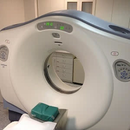
Peripheral arterial occlusive disease (PAOD) is a common condition associated with arterial wall degeneration mainly caused by arteriosclerosis. Diffuse progressive accumulation of arterial wall plaque results in luminal narrowing of the peripheral arteries and ultimately leads to organ ischaemia. Despite advances, challenges remain for less invasive imaging of PAOD using computed tomography (CT) angiography, according to an article published online in the journal Clinical Radiology.
CT is a useful noninvasive technique not only for the detection of luminal stenosis but also for plaque characterisation; however, particularly in patients with PAOD, extensive arterial calcification accumulates in the distal arteries. In such patients, the article says, accurate visualisation of the lumen with conventional CT is hampered by the artefacts caused by calcifications, as well as the lower resolution of CT than that of digital subtraction angiography (DSA).
"Recently, an ultra-high-resolution CT (UHRCT) system has been introduced, and it allows a more than twofold increase in spatial resolution. The high spatial resolution may be particularly beneficial in the visualisation of small vasculature," the article notes. "In combination with advances in post-processing, such as subtraction techniques, these developments may overcome some of the current challenges and allow for more detailed and noninvasive characterisation of PAOD."
UHRCT (Canon Medical Systems, Otawara, Japan) is a 160-row multidetector CT with superfine detector grids providing an effective detector element size as small as 0.25×0.25 mm and 1,792 detector channels. UHRCT also has a fine x-ray focus size as small as 0.4×0.5 mm. Controlled-orbit helical scanning, which synchronises the helical orbit of data acquisition and patient table feed is available for the subtraction technique.
"The 0.25-mm effective size of the detector element on UHRCT enables the visualisation of smaller structures more precisely. Indeed, compared to currently available CT systems, this system allows for extensive and more detailed visualisation of the distal vasculature," the article points out.
In particular, UHRCT is utilised in the noninvasive visualisation of the collateral arteries, pedal arch, and digital arteries. Evaluation of distal smaller arteries is very important in patients with severe limb ischaemia. Noninvasive visualisation of such smaller distal arteries, especially prior to invasive procedures, is needed to determine an adequate treatment strategy.
Subtraction CT is a technique for extracting the contrast-enhanced area (vascular lumen) similar to DSA. According to the article, a basic concept in subtraction CTA using controlled-orbit helical scanning is to scan slowly from the upper abdomen to the ankle and to maintain luminal radiopacity during the scan. In patients with significantly slow blood flow, this slow scanning approach remains faster than the analysis of their blood flow.
"Basic subtraction is effective when the object (patient) stands still; however, inconsistency between datasets can occur because of movements such as breathing, bowel motion, pulsation of vessels, and involuntary movement. Because subtraction CT is based on three-dimensional data, unlike DSA (which considers two-dimensional data), it is conceivable that such inconsistencies between two datasets may have substantially greater impact. Therefore, advanced post-processing is required for obtaining accurate subtracted images in CT," the article explains.
Alternative approaches to overcome some of the limitations of CT in the visualisation of calcifications and the small arteries of the lower vasculature include dual-energy CT and dynamic CT. Currently, both techniques are unavailable in combination with UHRCT, the article says.
"As UHRCT has only recently become available, no systematic studies have been conducted. Accordingly, while our initial experience is promising, its ability for visualising the entire lower extremity arterial circulation has not yet been established. Further evaluations are necessary to establish its full utility for diagnosis and decision-making in patients with PAOD," Ryoichi Tanaka, MD, PhD, of the Department of Radiology, Memorial Heart Center, Iwate Medical University (Morioka, Japan) and co-authors conclude.
Source: Clinical Radiology
Image Credit: Pixabay
References:
Tanaka R et al. (2018) Novel developments in non-invasive imaging of peripheral arterial disease with CT: experience with state-of-the-art, ultra-high-resolution CT and subtraction imaging. Clinical Radiology, Published online: 04 April 2018 DOI: https://doi.org/10.1016/j.crad.2018.03.002
Latest Articles
CT, peripheral angiography, Peripheral arterial occlusive disease, PAOD
Peripheral arterial occlusive disease (PAOD) is a common condition associated with arterial wall degeneration mainly caused by arteriosclerosis. Diffuse progressive accumulation of arterial wall plaque results in luminal narrowing of the peripheral arteri


