According to researchers, cardiac hybrid imaging with CT and nuclear stress testing is an excellent long-term predictor of adverse cardiac events like heart attacks in patients being evaluated for coronary artery disease (CAD). Hybrid imaging findings could help guide treatment decisions, such as whether or not a patient should have a revascularisation procedure such as bypass or angioplasty, the research team explains.
CAD is a leading cause of death and disability worldwide. The current gold standard for determining the percent of stenosis (or narrowing), due to plaque in a coronary artery, is invasive coronary angiography (ICA). However, the degree of stenosis on ICA is not always an accurate predictor of heart attack risk because it gives no information on perfusion, or the flow of blood into the heart muscle. Inadequate perfusion, also known as ischaemia, is a potential danger to the patient.
In comparison, cardiac hybrid imaging combines coronary computed tomography angiography (CCTA) and myocardial perfusion imaging with single photon emission tomography (SPECT) to provide information on both stenosis and perfusion. Although this approach has shown promise in studies focusing on short-term observations, information is lacking on long-term outcomes.
The research team looked at 428 patients who underwent hybrid imaging. During a median follow-up of 6.8 years, a total of 160 major adverse cardiac events, including 45 deaths, were observed in the final study population. Patients with matched findings – stenosis of 50 percent or more on CCTA with evidence of ischaemia on SPECT in the area of the heart to which the blocked vessel was supplying blood – had more than five times the risk of adverse events than those with normal findings. Patients with unmatched findings, or evidence of ischaemia but not in the area of the heart being fed by the stenotic artery, had three times the risk. Major adverse cardiac event rates were 21.8 percent for matched findings and 9.0 percent for unmatched – considerably higher than the 2.4 percent rate for normal findings.
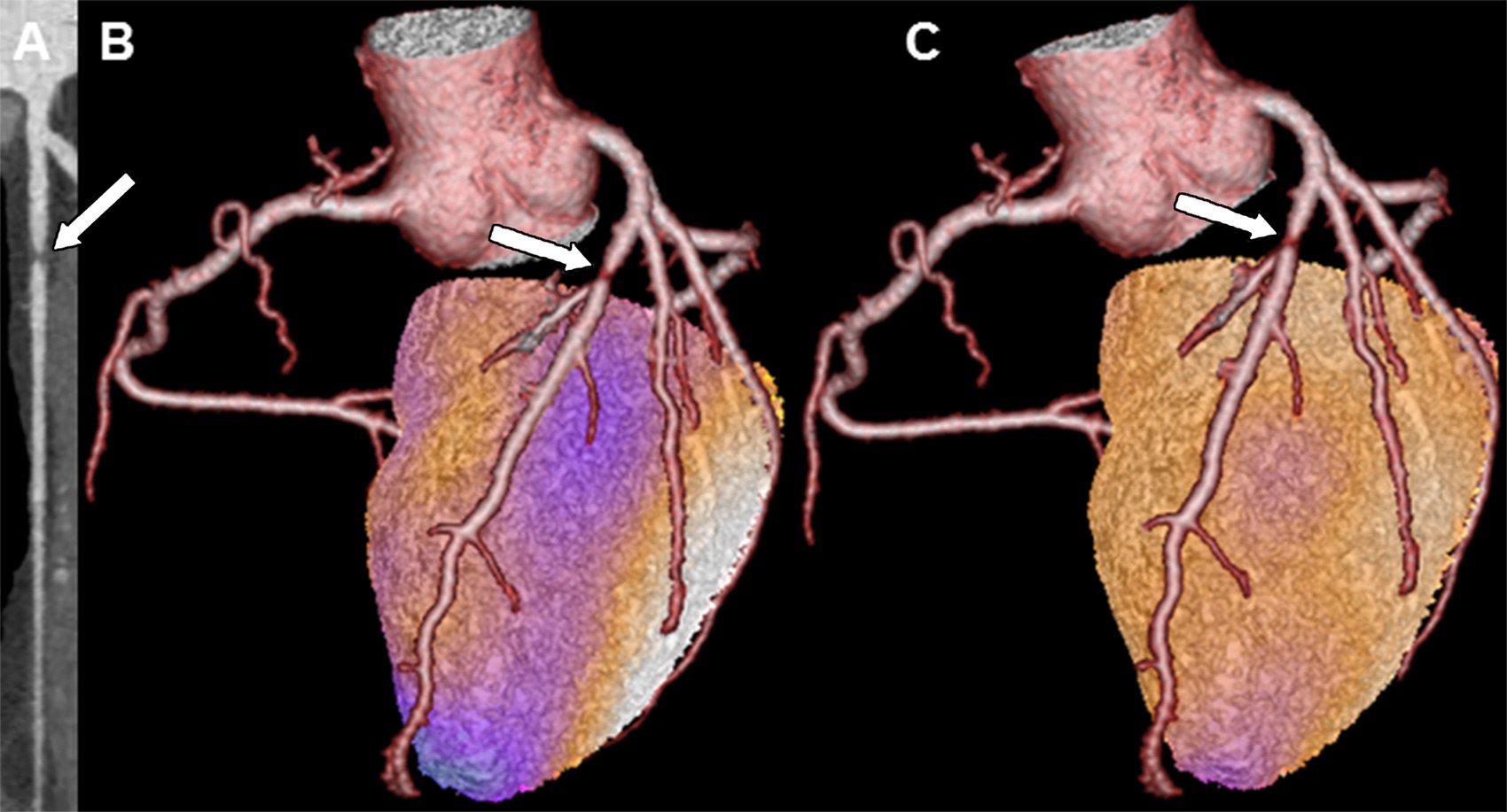
Figure 1. Example images in a patient with a matched cardiac hybrid finding (arrows). A, Multiplanar contrast material–enhanced coronary CT angiography reconstruction shows a stenosis of the proximal left anterior descending artery in a 62-year old man. B, C, Cardiac hybrid SPECT/coronary CT angiography images show a matched perfusion defect (arrow) in the territory served by the left anterior descending artery at, B, stress; this defect was reversible at, C, rest, indicating a stress-induced anterior ischaemia.
The results highlight the "prognostic importance of the comprehensive assessment provided by hybrid imaging,” said study co-author Philipp A. Kaufmann, MD, professor and chair of nuclear medicine, and director of cardiac imaging at University Hospital Zurich in Switzerland (pictured, top). “In a previous multicentre trial, with hybrid imaging we were able to see that about one in five patients should be revascularised in another coronary artery than originally planned."
The study findings support CCTA use for an initial, noninvasive evaluation of patients with known or suspected stable CAD. No additional imaging would be necessary if the results were normal. If a lesion was evident, then clinicians could employ a nuclear scan to assess ischaemia and take advantage of both modalities by fusing the results together to make a hybrid image.
“The strategy of direct referral to invasive coronary angiography without noninvasive imaging is obsolete,” Dr. Kaufmann emphasised. “Even after documenting coronary artery disease with coronary CT angiography, we need further noninvasive evaluation before deciding upon revascularisation versus medication.”
The research team hopes to run a trial to demonstrate that hybrid imaging can have a positive impact on patient outcomes.
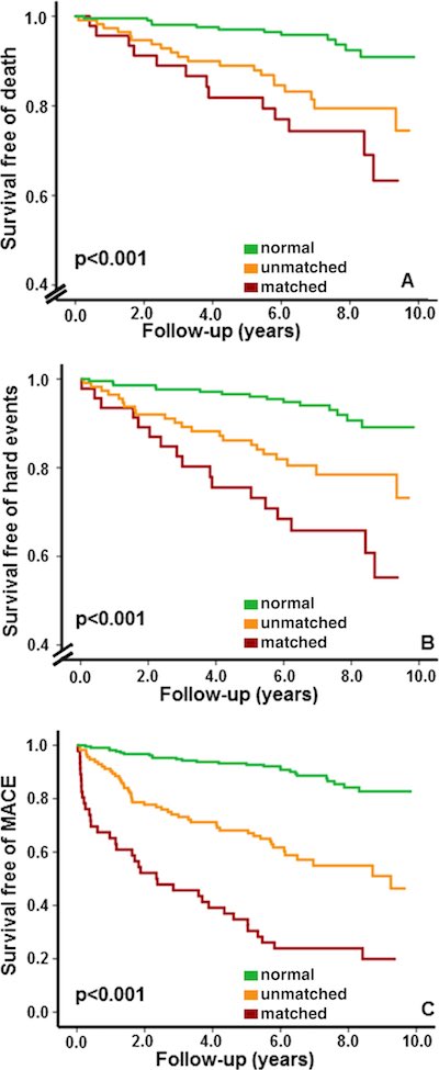
Figure 2. Kaplan-Meier survival curves show prognostic value of cardiac hybrid imaging
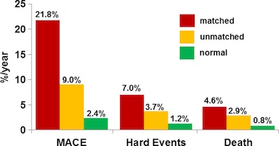
Figure 3. Bar graph shows annual event rates
corresponding to cardiac hybrid imaging finding. MACE = major adverse
cardiac event. Matched (findings) = stenosis of 50 percent or greater
(at coronary CT angiography) with ischemia (at SPECT) in subtended
territory, unmatched (findings) = coronary CT angiography and/or SPECT
findings in unrelated territories
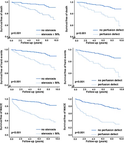
Figure 4. Kaplan-Meier survival curves show prognostic value of stenosis of 50 percent or greater alone and perfusion defect alone. Left: Patients with a stenosis of 50 percent or greater alone and right: a perfusion defect alone had worse long-term outcomes than patients with no stenosis and no perfusion defect, respectively
Image Credit: RSNA
References:
Pazhenkottil AP, Benz DC, Gräni C, Madsen MA, Mikulicic F, von Felten E, Fuchs TA, Hirt Moch B, Stehli J, Lüscher TF, Gaemperli O, Buechel RR, Kaufmann PA (2018) Hybrid SPECT perfusion imaging and coronary ct angiography: long-term prognostic value for cardiovascular outcomes. Radiology, Jul 3. doi: 10.1148/radiol.2018171303
Latest Articles
CT, heart attacks, cardiac hybrid imaging, nuclear stress testing
According to researchers, cardiac hybrid imaging with CT and nuclear stress testing is an excellent long-term predictor of adverse cardiac events like heart attacks in patients being evaluated for coronary artery disease (CAD). Hybrid imaging findings cou