In a study presented at the annual meeting of the Radiological Society of North America (RSNA) in Chicago, researchers using artificial intelligence have developed a deep learning algorithm that may lead to the reduction of the gadolinium dose used in contrast material for MRI exams.
“There is concrete evidence that gadolinium deposits in the brain and body,” said lead author of the study Enhao Gong, researcher at Stanford University. “While the implications of this are unclear, mitigating potential patient risks while maximising the clinical value of the MRI exams is imperative.”
You may also like: Radiologists do not routinely report gadolinium deposition findings
Results of recent studies have shown that trace amounts of the heavy metal gadolinium used in contrast material to enhance images on MRI remain in the body of people who undergo exams. Radiologists have since been active in working towards increasing patient safety while not compromising the important information gadolinium contrast MRI scans provide.
To help achieve this goal Dr. Gong and his research colleagues at Stanford have been investigating the potential of applying deep learning- a sophisticated artificial intelligence method that teaches computers to learn by feeding them data examples. Using convolutional neural networks, the computer can recognize images and find subtle distinctions within the imaging data that a human expert might not be able to detect.
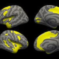
The researchers collected three sets of images from 200 patients who had received contrast-enhanced MRI exams for various indications to train the deep learning algorithm. The three sets of images from each patient were pre-contrast scans performed before contrast administration and referred to as the zero-dose scans, lose-dose scans after 10 percent of the gadolinium dose administration and full dose scans at 100 percent of the administered dose.
The deep learning algorithm learned to distinguish the full-dose scans from the zero-dose and low-dose images, and neuroradiologists assessed the images for contrast enhancement and quality.
The results found that MR image quality was not significantly different between full-dose contrast enhanced images and the low-dose images that were enhanced by the artificial intelligence algorithm. In addition the research results also showed the potential to create the equivalent of full-dose contrast enhanced MR images without using any contrast agent.
“Low-dose gadolinium images yield significant untapped clinically useful information that is accessible now by using deep learning and AI,” said Dr. Gong. He also added that these results demonstrate AI’s potential to dramatically reducing gadolinium dose without sacrificing the diagnostic quality of the images.
The next step after showing that deep learning potential is technically possible, Dr. Gong and his colleagues want to further study the method in clinical use where it will ultimately find a home. This future research will include evaluating the algorithm using a broader range of MRI scanners and different contrast agents.
“We’re not trying to replace existing imaging technology,” Dr. Gong said. “We’re trying to improve it and generate more value from the existing information while looking out for the safety of our patients.”
Image Credit: RSNA
References:
Latest Articles
MRI, breast cancer, breast screening, AI, Stanford University, #RSNA, Artificial Intelligence, gadolinium-based contrast agents, deep learning, #RSNA18
In a study presented at the annual meeting of the Radiological Society of North America (RSNA) in Chicago, researchers using artificial intelligence have developed a deep learning algorithm that may lead to the reduction of the gadolinium dose used in con