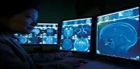
Researchers at UC San Diego School of Medicine have tested a new magnetic resonance imaging (MRI)-based technique to better detect and evaluate nonalcoholic fatty liver disease (NAFLD) in children. The new method is known as magnitude-based MRI, which was previously developed by researchers in the UC San Diego Liver Imaging Group, to estimate liver proton density fat fraction (PDFF), a biomarker of liver fat content.
The investigators found a strong correlation between the amount of liver fat as measured by the new MRI technique and the grade of liver fat determined by pathology. Their findings are published in Hepatology.
NAFLD is characterised by large droplets of fat in at least five percent of a child’s liver cells. Traditionally, NAFLD is diagnosed by a gastroenterologist in consultation with a pathologist, who examines the patient’s biopsied liver tissue under a microscope. The presence and severity of liver fat is graded by the pathologist as none, mild, moderate or severe, based on the percentage of liver cells that contain fat droplets.
“Currently, diagnosis of NAFLD requires a liver biopsy, which is not always available or performed. This leads to both misdiagnosis and missed diagnoses, hampering patient care and progress in clinical research,” said Jeffrey B. Schwimmer, MD, professor of clinical paediatrics at UC San Diego, director of the Fatty Liver Clinic at Rady Children’s Hospital-San Diego and the first author of the study. “Thus, a noninvasive method for diagnosing and/or evaluating NAFLD has the potential to impact millions of children.”
In this study (MRI Rosetta Stone Project), Dr. Schwimmer and colleagues compared the new MRI technique to the standard liver biopsy method of assessing fat in the liver. To do this, the team enrolled 174 children who were having liver biopsies for clinical care. For each patient, the team performed MRI-estimated PDFF and compared the results to the standard pathology method of measuring fat on a liver biopsy. Their analysis showed a strong correlation between the two techniques in assessing fat in the liver.
Notably, the correlation was influenced by both the patient’s gender and the amount of scar tissue in the liver. The correlation between the two techniques was strongest in females and in children with minimal scar tissue.
“Existing techniques for measuring liver fat are dependent upon the individual scanner and the centre at which the measurements were made, so they cannot be compared directly,” explained Claude B. Sirlin, MD, professor of radiology at UC San Diego and senior author of the study. “By comparison, PDFF is a standardised marker that is reproducible on different scanners and at different imaging centres. Thus, the results of the current study can be generalised to the broader population.”
Depending on how the new MRI technology is used, it could correctly classify between 65 and 90 percent of children as having or not having fatty liver tissue.
“We are especially excited about the promise of the technology for following children with NAFLD over time. However, further refinements will be needed before this or any other MRI technique can be used to diagnose NAFLD in an individual child,” Dr. Schwimmer said.
Obesity and diabetes are risk factors for NAFLD, which affects between 5 and 8 million children in the United States. Doctors are concerned about NAFLD in children because it can lead to hepatitis, liver scarring, cirrhosis and liver cancer.
Source: UC San Diego School of Medicine
Image Credit: UC San Diego School of Medicine
Latest Articles
MRI, diabetes, obesity, pathology, fatty liver, NAFLD, hepatitis
Researchers at UC San Diego School of Medicine have tested a new magnetic resonance imaging (MRI)-based technique to better detect and evaluate nonalcoholi...