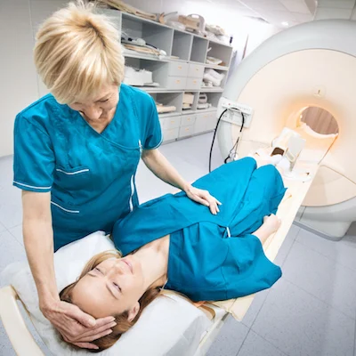
One of the major problems which is commonly attributed to affect patient outcomes is the failure to follow-up diagnostic imaging in time. It is critical to follow-up diagnostic imaging findings in order to have access to and receive high quality and vital care. Patient location at the time of undergoing imaging is directly related with how likely a patient is of completing the necessary follow-up imaging for lesions with indeterminate malignant potential, according to a report published in the Journal of the American College of Radiology (JACR ).
The threat to patient safety and quality of care continues to grow as a result of failing to complete follow-ups of abnormal findings especially in the cases presenting with malignant potential- leading to delayed or missed crucial diagnoses. To try and address this significant issue, healthcare systems are increasingly focusing on dealing with any factors at the clinical level that may influence imaging findings follow-up by reviewing results notification and efficient communication between radiologists, referring physicians and imaging ordering providers. In addition to the importance of targeting interventions at individual providers, researchers aimed to determine how broader healthcare system factors may play a role in the failure to complete recommended imaging follow-ups.
You may also like: Is walk-in CT better than an appointment system?
Healthcare practices and delivery processes are vastly different in reference to emergency department (ED), inpatient and outpatient settings. Nevertheless, all three departments share the common problem of failed radiology exam follow-ups that often lead to negative patient outcomes. In this report, researchers consider that empirical evidence concerning the correlation between patient location at the time of imaging and the relevant imaging follow-up provides valuable insight that healthcare providers can use to understand which of their care processes they can prioritise in order to increase the care quality they provide within the different departments.
The research team ventured to examine this missing link in information by evaluating the relationship between where the patient is located at time of imaging and the subsequent completion of the necessary imaging follow-up for findings in the abdomen with indeterminate malignant potential. They set out on a basis that findings from exams performed in the ED were less likely to be followed-up in comparison with the ones performed while the patient was in the hospital or in outpatient settings. In order to determine the possible clinical implications of imaging follow-up, the researchers estimated the progression rate of the findings to the ones that presented suspicions for malignancy.
The team identified all abdominal ultrasound (US), CT, and MR exams performed at their institution from July 1, 2013, to January 31, 2014. Mass lesions identified in the liver, adrenal glands, kidneys, and pancreas were assigned mandatory standardised assessment categories, which group lesions as benign, indeterminate, or suspicious for malignancy; biopsy-proven known malignancy; or incompletely evaluated with the performed modality. Using this system, they identified exams presenting lesions indeterminate for malignancy, which they then defined as those categorised either as indeterminate or as incomplete. Recommendations for imaging follow-up based on the patient’s clinical context were given for all the lesions categorised as indeterminate or incompletely evaluated.
For each unique patient, they assigned an index examination as the first during the study period with an indeterminate lesion in any of the four organs of interest. Then defined relevant imaging follow-up as subsequent outpatient abdominal cross-sectional imaging of the corresponding organ within 13 months of the index examination (to accommodate a 12-month follow-up period and 1-month grace period). Progression from an indeterminate lesion to a suspicious lesion on imaging or a biopsy-proven malignant lesion was determined using categories assigned to the organ of interest on follow-up imaging examinations within 395 days of the index examination.
From 1,142 identified index examinations, they excluded the ones from patients who either died within 13 months of the index examination or who received repeat imaging in the ED or inpatient settings before any subsequent outpatient imaging and had the initial indeterminate lesion downgraded to benign or upgraded to one suspicious for or proven to be malignancy. This process produced a final sample consisting of 1,032 eligible index examinations.
The predictor of interest—patient location at the time of index examination—was classified as either an outpatient, ED, or inpatient setting. They analysed the modality of the index examination, along with characteristics of the interpreting radiologist (gender, years of experience) and ordering providers (clinical specialty, physician versus non-physician provider), as well as data about patient sociodemographics (age, gender, race, median income, and percentage of individuals under the federal poverty line in the zip code of residence) and clinical conditions (existing cancer diagnosis, other clinical comorbidities).
The study found that 70% of exams were performed in the outpatient setting compared with 13% and 16% in the inpatient and ED settings. Approximately half (53%) were interpreted by male radiologists, and most (75%) were interpreted by radiologists with more than 10 years of professional experience. Most exams were ordered by clinicians in the fields of general and subspecialty internal medicine (50%) or general and subspecialty surgery (31%). The majority of exams were also ordered by physician (83%) rather than non-physician (17%) providers.
Patient location is correlated with the likelihood of completing relevant follow-up imaging for lesions with indeterminate malignant potential. Future work should evaluate delivery processes, care continuity, and other health system–level factors related to patient location and care setting as well as their effects on appropriate follow-up of these findings. Doing so would support efforts to improve appropriate follow-up imaging and address health care disparities.
- Patient location at time of imaging is associated with the likelihood of completing relevant follow-up imaging for lesions with indeterminate malignant potential.
- Health systems could improve follow-up for imaging done in ED and inpatient settings by implementing system-level changes that span provider types, service lines, and sites of care.
- Efforts should be pursued with recognition of the disparities in follow-up imaging for black patients and individuals residing in lower-income neighbourhoods.
- The relatively low rate of progression of imaging findings from indeterminate to suspicious for or known malignancy suggests that follow-up may be deferred in many cases to the outpatient setting rather than systematically prioritised during acute ED or inpatient stays.
It is very important for health systems to identify and address differences in imaging follow-ups based on the care setting so that they can adjust organisational strategies related to healthcare quality improvement.
The researchers found that relevant follow-up occurred in only half of all examinations, a result that underlines the need to increase efforts to redesign care processes to improve imaging follow-up for findings with indeterminate malignant potential.
In addition, they determined that follow-up varies based on patient location which supports the notion that health system–level factors influence completion of relevant follow-up. In particular, follow-up for exams performed in the ED was much lower than that for exams performed in the outpatient setting. However, follow-up was also lower for examinations performed in the inpatient setting compared with the outpatient setting.
These results reinforce common issues with communication about and monitoring of test follow-up during transitions after hospital discharge and potentially ED visits, as well as other elements that increase the probability that results from ED and inpatient exams are missed or incompletely followed up. Radiology tests may be obtained in the outpatient setting to evaluate urgent or non-urgent health issues, whereas ED and inpatient imaging exams are more commonly used to help evaluate acute complaints. Patients are more likely, therefore, to accrue a larger total number of exams and utilise more imaging modalities across an ED or inpatient stay—each of which must be noted, communicated, and followed up after the acute care encounter—than they might as part of a single outpatient evaluation.
Health systems could implement several system-level changes to improve follow-up for imaging performed in ED and inpatient settings. For example, pre-discharge checklists could be used to ensure that actionable findings from imaging exams performed during a hospital admission are flagged and addressed. Similar interventions could be used in the ED and paired with other efforts, such as ED-made appointments or pre-discharge questionnaires that screen for risk factors of failed adherence or follow-up. Health systems that use either electronic health records or physical discharge coordinators, these requirements could be 'built-into' processes through electronic entry prompts or mandatory manual staff procedures. Codifying processes in the ED or hospital to record information about patients’ primary care providers could also facilitate communication of discharge summaries, checklists, and actionable tests to follow-up.
These checks align well with evidence about the benefits of structured inter shift hand-offs and could improve communication at the time of unit transfer (eg, ED to inpatient, across different inpatient services and units) and ensure that receiving care teams are aware of findings with indeterminate malignant potential. Doing so systematically across providers in certain service lines could further improve imaging follow-up, a notion supported by our finding that examinations ordered by surgical providers in our sample were less likely to receive relevant follow-up.
Third, the report found evidence of disparities in follow-up imaging for black patients and individuals residing in lower-income neighbourhoods. These results are consistent with existing evidence of disparities in receipt of scheduled outpatient imaging (eg, mammography screening, CT and MRI studies). The study extends this body of evidence by demonstrating the issue within the context of patient location during imaging and the results underscore the need to connect system-level strategies with those focused on patient engagement and access.
Finally the study found that the progression of indeterminate findings to more worrisome lesions was relatively rare and consistent across patient location. Although this result should be verified across institutions in future work, it provides some evidence that the exact timing of follow-up may not be critical and that some imaging findings could be deferred to outpatient follow-up rather than systematically prioritised during acute ED or inpatient stays.
The report notes that this study possesses limitations. First, although they adjusted analysis for factors that are plausibly associated with completion of relevant imaging follow-up, some others may not have been included. Second, results from this retrospective analysis should be further studied using prospective design. Nonetheless, as an exploratory analysis, the study reveals associations that merit future work. Third, the analysis was performed using index and follow-up examinations conducted at a single academic institution. Although this design limits overall generalisability and the ability to observe follow-up care at outside organisations, the approach also has “real-world” relevance given that quality improvement efforts are implemented at the organisational level.
Sources: JACR
Image credit: iStock
References:
Latest Articles
Radiology, CT scans, MR, Cancer imaging, MRI, Health Systems, Follow-up Imaging, Care Delivery, follow-up
One of the major problems which is commonly attributed to affect patient outcomes is the failure to follow-up diagnostic imaging in time. It is critical to follow-up diagnostic imaging findings in order to have access to and receive high quality and vital


