An innovative new technique developed by UNC researchers could enable doctors to visualise tumours without using radiation in a more cost effective way.
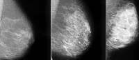
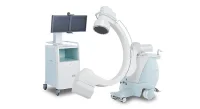
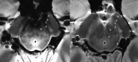
Compared to X-rays, CT scans, and MRIs, ultrasound not only provides an alternative which is less expensive, but more importantly, it is a radiation-free technology for detecting and monitoring cancer. Still, due to limited clarity and resolution issues, its use has been limited.
Providing a way around these limitations, a team of researchers from the UNC School of Medicine has pioneered a new method by combining ultrasound with a contrast agent composed of tiny bubbles. These are capable of pairing with an antibody that many cancer cells produce at higher levels than normal cells do.
Through this binding process to the protein SFRP2, the microbubble contrast agent greatly enhances the resolution and tumour-detecting ability of scans produced by ultrasound. Publishing their findings in PLOS ONE, UNC Lineberger Comprehensive Cancer Center members Nancy Klauber-Demore, MD, professor of surgery and Paul Dayton, PhD, professor of biomedical engineering, described how they succeeded in visualising lesions created by angiosarcoma, a malignant cancer that develops on the walls of blood vessels.
As Klauber-DeMore explained, the SFRP2-moleculary targeted contrast agent showed specific visualisation of the tumor vasculature while there was no visualisation of normal blood vessels. This result suggests to the authors that the contrast agent may help distinguish malignant from benign masses found on imaging.
Based on their pioneering discovery that angiosarcoma cells produce an excess of SFRP2, Klauber-DeMore’s team concentrated on ways to utilise the protein in order to better monitor the progress of the cancer within blood vessels. Through the use of a mouse model, the researchers delivered the microbubble contrast agent via intravenous injection and tracked it using ultrasound.
Due to the fact that SFRP2 is expressed in many cancers such as breast, colon, pancreas, ovarian, and kidney tumours, it is hoped that this method could potentially be efficient on a wide range of cancer types. The team’s next objective is to determine how well the technique works with these other tumor types, as well as to study its effect on breast cancer.
Previous research has indicated that SFRP2 levels in tumours increase as tumours develop, hence Klauber-DeMore and her colleagues will also investigate whether the technique can be used to track tumour growth, making it useful in monitoring patient response to chemotherapy. Additionally, they will look into whether very small tumors can also be detected and visualised this way.
Ultrasound is less expensive than commonly used imaging methods, hence making it a cost effective method in cancer treatments, and since an ultrasound machine is more portable than other imaging devices, it may be useful in providing treatment in rural areas.
Source: UNC
18 February 2014
Latest Articles
Cancer, X-ray, MRI, Ultrasound, Tumours, breast cancer, Visualisation, CT scans, cells, proteins, colon cancer
An innovative new technique developed by UNC researchers could enable doctors to visualise tumours without using radiation in a more cost effective way....