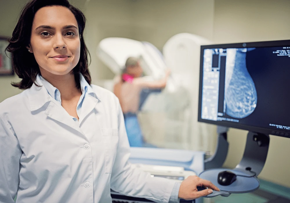
Breast cancer remains a significant health concern, particularly for women with dense breast tissue. High mammographic density not only elevates the risk of breast cancer but also complicates the detection process, often leading to missed diagnoses. Mammography, the standard screening tool, has reduced sensitivity in women with dense breasts, which can result in undetected cancers. Supplemental imaging techniques, such as breast MRI, have been proposed as an adjunct to mammography to improve cancer detection rates in this high-risk population. A systematic review recently published in the Journal of Breast Imaging explores the effectiveness of supplemental breast MRI in detecting breast cancer in women with dense breasts, summarising key findings from a systematic review and meta-analysis.
The Impact of Dense Breasts on Mammographic Sensitivity
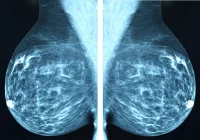
Dense breast tissue is composed of less fat and more fibroglandular tissue, which appears white on a mammogram—the same colour as potential malignancies. This similarity can obscure the presence of tumours, making it difficult to distinguish between healthy dense tissue and cancerous growths. Consequently, mammograms are less sensitive in detecting cancer in dense breasts, increasing the likelihood of interval cancers, which are cancers diagnosed between regular screening intervals. This masking effect necessitates alternative screening strategies to ensure early detection, where supplemental breast MRI has shown promise.
Effectiveness of Supplemental MRI in Cancer Detection
Supplemental MRI screening after a negative mammogram has demonstrated a significantly higher cancer detection rate (CDR) than mammography alone. The meta-analysis reviewed studies involving over 21,000 women, revealing that MRI detected approximately 16.6 cancers per 1,000 exams in initial (prevalent) screenings and 6.8 cancers per 1,000 exams in subsequent (incident) screenings. This increased detection rate is significant in women with dense breasts, where mammograms may fail to identify malignancies. The review also highlighted that MRI is especially effective in detecting mammographically occult cancers, with a sensitivity of 98.4% in women who had previously received a negative mammogram.
Considerations and Challenges in Implementing Supplemental MRI
While the benefits of supplemental MRI are evident, several considerations and challenges are associated with its widespread adoption. One significant factor is the increased recall rate associated with MRI, leading to more follow-up tests and potentially unnecessary biopsies. This can contribute to patient anxiety, increased healthcare costs, and the physical and emotional burden of additional procedures. Moreover, the availability of MRI and the costs associated with it may limit its use, especially in lower-resource settings or for women who do not fall under high-risk categories, such as those with a strong family history of breast cancer or known genetic mutations.
Conclusion
Supplemental breast MRI represents a valuable tool in the early detection of breast cancer for women with dense breasts, significantly improving the cancer detection rate beyond what mammography can achieve alone. However, its implementation must be carefully considered, balancing the benefits of early detection with the potential drawbacks of increased recall rates and costs. As research continues and MRI technology advances, it may become more feasible and widely accepted as a routine supplement to mammography for women with dense breasts, particularly as guidelines evolve to reflect the growing body of evidence supporting its use. Future studies should focus on refining MRI protocols to minimise false positives and on exploring cost-effective strategies to make this life-saving technology accessible to a broader population.
Source: Journal of Breast Imaging
Image Credit: iStock


