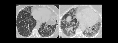
Hypersensitivity pneumonitis (HP) is a complex and often elusive form of interstitial lung disease (ILD), primarily triggered by repeated exposure to airborne antigens. The diagnosis of HP is challenging due to the heterogeneous nature of its clinical and radiologic presentations, which often overlap with other forms of ILD. Accurate and timely diagnosis is essential for optimising patient outcomes, as it allows for the prompt initiation of treatment and the avoidance of further exposure to the causative antigen. The introduction of updated classification schemes by the American Thoracic Society, Japanese Respiratory Society, and Asociación Latinoamericana del Tórax (ATS/JRS/ALAT) and the American College of Chest Physicians (ACCP) has provided clinicians with new tools to diagnose HP using thin-section chest CT scans. This article explores the diagnostic performance of these two classifications, highlighting their strengths, limitations, and implications for clinical practice.
ATS/JRS/ALAT Classification: Emphasis on Radiologic and Histologic Assessments
The ATS/JRS/ALAT classification for HP focuses heavily on the presence or absence of fibrosis and the distribution of radiologic findings. This classification system categorises CT scan findings into typical HP (tHP), compatible HP (cHP), and indeterminate HP (iHP). Diagnosing tHP requires a combination of small airway disease manifestations and parenchymal infiltration or fibrosis, with a diffuse distribution of findings expected in nonfibrotic HP. In fibrotic HP, the findings should be diffusely distributed with mid-lung predominance or relative basilar sparing. The ATS/JRS/ALAT system offers high specificity, particularly in low-prevalence settings, making it a robust tool for ruling out HP when radiologic features are absent. However, its stringent criteria may result in lower sensitivity, especially in cases where radiologic findings do not perfectly align with the typical patterns outlined by the guidelines.
ACCP Classification: A More Lenient Approach with Higher Sensitivity
The ACCP classification adopts a more lenient approach compared to ATS/JRS/ALAT, allowing for a high-confidence diagnosis of tHP with less stringent radiologic criteria. This classification does not emphasise the distribution of abnormalities as strongly as ATS/JRS/ALAT and permits a tHP diagnosis based on diffuse centrilobular nodularity alone, provided that alternative diagnoses are excluded. The ACCP classification demonstrates greater sensitivity, making it particularly useful in high-prevalence settings with a higher likelihood of HP. This leniency, however, may lead to a lower positive predictive value (PPV) in low-prevalence environments, where overdiagnosis could occur due to the broader criteria.
Comparative Performance: The Impact of Disease Prevalence
The diagnostic performance of ATS/JRS/ALAT and ACCP classifications varies significantly with disease prevalence. In low-prevalence settings, the ATS/JRS/ALAT classification outperforms the ACCP guidelines in terms of accuracy and PPV. Its higher specificity ensures that patients without HP are less likely to be misdiagnosed, which is crucial in environments where HP is rare. Conversely, in high-prevalence settings, the ACCP classification shows greater accuracy and negative predictive value (NPV), making it more reliable in confirming HP diagnoses. The study also found a discordance rate of 21% between the two classifications, with most disagreements occurring between the tHP and cHP categories. These discrepancies underscore the importance of considering disease prevalence and the specific clinical context when selecting a diagnostic approach.
Conclusion
The diagnosis of hypersensitivity pneumonitis remains challenging, particularly due to the variability in disease presentation and prevalence. The ATS/JRS/ALAT and ACCP classifications offer complementary strengths; the former provides greater specificity in low-prevalence settings, while the latter offers higher sensitivity in high-prevalence environments. Clinicians must carefully consider the prevalence of HP in their patient population when choosing between these classification schemes to optimise diagnostic accuracy and patient outcomes. Further research is needed to refine these guidelines and explore their impact on clinical decision-making and prognosis.
Source & Image Credit: Radiology: Cardiothoracic Imaging


