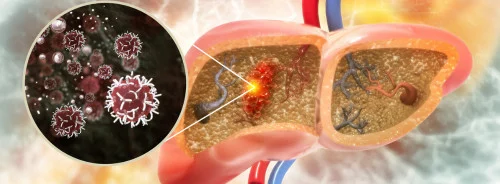
Hepatocellular carcinoma (HCC) remains one of the leading causes of cancer-related deaths globally, with mortality rates rising steadily. Accurate mortality risk prediction is a cornerstone for optimising treatment and management strategies in patients diagnosed with HCC. Traditional scoring systems rely on simplistic models that consider limited clinical factors, often neglecting the rich data provided by advanced imaging modalities. However, the advent of machine learning (ML) techniques in radiomics has opened new avenues for predicting patient outcomes with greater precision. A recent article from European Radiology explores the application of a machine-learning-based prognostic tool developed to predict mortality in HCC patients, utilising clinical data and radiomic features extracted from magnetic resonance imaging (MRI).
Development of a Machine Learning-Based Prognostic Model
Traditional methods for predicting HCC outcomes, such as the Child-Pugh score or Barcelona Clinic Liver Cancer (BCLC) staging, use a narrow set of variables, primarily clinical and tumour size measurements, which may not capture the complete picture of the disease. These systems are limited by their reliance on one-dimensional tumour size measurements and subjective threshold-based risk stratification. In contrast, the new ML-based model takes advantage of the full three-dimensional volume of liver imaging data, using radiomic features extracted from multiphasic MRI scans.
The radiomic data is obtained through automated liver segmentation, ensuring reproducibility and removing variability introduced by human interpretation. This fully automated framework reduces processing time, completing the analysis in just over a minute per patient, making it practical for clinical settings. The model uses a random survival forest (RSF), a machine-learning method that combines clinical and radiomic features to predict overall survival (OS). This approach considers both the complexity of the liver tumour and the underlying liver disease, capturing crucial information such as tumour texture and intensity variations across different imaging phases.
Validation and Performance of the Model
The ML-based model was developed using a dataset of 555 treatment-naïve HCC patients who underwent multiphasic MRI at diagnosis. After development, the model was independently validated with a subset of the patients. The model's performance was evaluated using Harrell’s C-index, which measures the ability of the model to predict outcomes correctly. The ML-based model outperformed conventional staging systems, achieving a C-index of 0.8503 in the development cohort and 0.8234 in the validation cohort, indicating its robustness.
Additionally, the model successfully stratified patients into low, intermediate, and high-risk groups. Each of these groups demonstrated significantly different survival times, confirming that the predictions were not only accurate but clinically meaningful. This risk stratification is valuable in guiding treatment decisions, as patients in the high-risk group might benefit from more aggressive treatment or closer monitoring, while low-risk patients could avoid overtreatment.
Comparison with Traditional Staging Systems
While the ML-based model showed superior performance, comparing it with conventional staging systems is essential to highlight its advantages. The Child-Pugh, BCLC, and the Hong Kong Liver Cancer (HKLC) staging systems are commonly used but have certain limitations. These traditional models rely on tumour size measurements, which do not always accurately reflect tumour viability or growth potential. Furthermore, they lack the depth of quantitative data provided by radiomics.
The ML-based model’s use of imaging biomarkers, such as texture analysis and intensity patterns, offers a more nuanced view of the tumour’s behaviour, which is crucial for predicting outcomes. It is essential for patients with smaller tumours or early-stage HCC, where conventional models might underestimate risk. The ML model’s ability to incorporate this wealth of data translates into more personalised and precise risk assessments.
Furthermore, traditional systems classify patients into rigid risk categories based on predefined cut-off values. This can result in misclassification, especially for patients whose disease characteristics fall on the border of these thresholds. The ML-based model, in contrast, uses data-driven cut-off points for stratification, making the classification more flexible and reflective of each patient’s unique disease profile.
Clinical Implications and Future Directions
Integrating machine learning into radiomics for HCC prognosis presents a promising advancement in personalised medicine. The ML-based model can significantly improve the accuracy and efficiency of mortality risk predictions by utilising routinely available clinical data and MRI scans. This improvement has practical implications in clinical settings, particularly for tumour boards where decisions about treatment strategies are made. With a more accurate prediction model, clinicians can tailor follow-up strategies to the patient's individual needs, ultimately improving outcomes.
This ML framework could be further enhanced by incorporating longitudinal data from different time points in a patient’s treatment journey, allowing for dynamic adjustments to risk predictions. Expanding the model to include external datasets from multiple institutions could help generalise its applicability across diverse patient populations and imaging protocols.
However, some limitations should be acknowledged. The current model is based on retrospective data from a single institution, and prospective multi-centre studies are needed to validate its generalisability. Furthermore, while the model provides excellent predictive accuracy, it does not yet account for the effects of different treatment modalities on survival outcomes. Incorporating treatment data into future model iterations could further refine its prognostic capabilities.
Conclusion
The application of machine learning in radiomics represents a significant leap forward in the prognostication of HCC. By leveraging automated image processing and advanced statistical models, the ML-based framework provides more accurate and personalised mortality risk predictions than conventional staging systems. This technology promises to enhance clinical decision-making and improve outcomes for patients with HCC, particularly as the model continues to evolve and incorporate additional data sources. As the field of radiomics grows, integrating AI-driven tools into routine clinical practice could revolutionise the management of liver cancer, ensuring that patients receive the most appropriate care based on their individual risk profiles.
Source Credit: European Radiology
Image Credit: iStock


