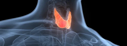
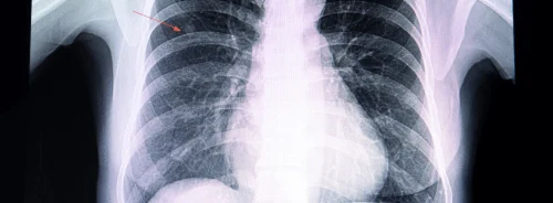
Thyroid cancer, particularly papillary thyroid cancer (PTC), has seen a global rise in incidence, making accurate and early diagnosis more crucial than ever. One of the critical aspects of thyroid cancer diagnosis is identifying lymph node metastasis (LNM), which significantly affects treatment plans and patient outcomes. While ultrasound (US) has long been the preferred imaging method for preoperative evaluation of thyroid cancer, its efficacy in detecting LNM is limited. As medical technology advances, radiomics, a technique that extracts quantitative data from medical images, has emerged as a potential game-changer. The combination of radiomics with ultrasound offers new possibilities for non-invasive, accurate diagnosis of LNM in thyroid cancer patients. However, the effectiveness and practicality of this integration are still under investigation. A recent article in Academic Radiology explores the utility of ultrasound-based radiomics for discerning lymph node metastasis in thyroid cancer and its benefits, challenges, and future potential.
Ultrasound-Based Radiomics: An Emerging Diagnostic Tool
Ultrasound is widely used to diagnose thyroid cancer due to its ability to provide real-time imaging and accessibility. However, its sensitivity, particularly in detecting LNM in the central cervical region, is suboptimal, leading to the frequent need for invasive diagnostic procedures such as fine needle aspiration biopsy (FNA) and preventive lymph node dissection (LND). This is where radiomics steps in. Radiomics transforms standard medical images into high-dimensional data, capturing details beyond the human eye's discernment, such as the tissues' texture, shape, and intensity features. These extracted features can be analysed using machine learning models to predict clinical outcomes like LNM.
Integrating radiomics with ultrasound aims to enhance the diagnostic accuracy of LNM detection. By combining clinical data and ultrasound-derived radiomic features, clinicians can better predict the likelihood of lymph node involvement in thyroid cancer patients, reducing the need for invasive procedures and improving preoperative planning. However, as research shows, while ultrasound-based radiomics holds promise, the current models exhibit variable accuracy, and some challenges need to be addressed for this technology to become a standard diagnostic tool.
Performance of Ultrasound-Based Radiomics in LNM Diagnosis
The performance of radiomics models for LNM detection in thyroid cancer has been the subject of extensive research. A recent meta-analysis of studies on ultrasound-based radiomics for thyroid cancer LNM found that models based solely on radiomic features tend to outperform those based only on clinical data. Specifically, models built from radiomic features had higher sensitivity and specificity, making them more reliable for identifying lymph node metastasis.
However, despite these promising results, the models are not without limitations. Combining clinical features with radiomics did not significantly improve diagnostic accuracy, suggesting that some clinical variables might already be reflected in the radiomic features. The synergy between clinical data and radiomic characteristics is still not fully understood, and more studies are needed to refine these models. In particular, there is a need for standardised protocols in image acquisition and feature extraction, as variability in these processes can affect the performance of radiomics models. Moreover, the high complexity of radiomics data presents a challenge in preventing overfitting of models, which could lead to less reliable predictions when applied in diverse clinical settings.
Challenges and Limitations in Implementing Radiomics
While ultrasound-based radiomics shows great potential for improving thyroid cancer diagnosis, several obstacles hinder its widespread adoption. One of the key challenges is the standardisation of imaging protocols. Different ultrasound machines, settings, and techniques can produce varying image qualities, affecting the consistency and reproducibility of radiomics models. Moreover, the segmentation of regions of interest (ROI) in the ultrasound images is often done manually, which introduces human variability. Automated or semi-automated segmentation tools could help mitigate this issue, but their development is still in progress.
Another challenge lies in the complexity of the radiomics models themselves. Radiomics generates many features from the images, many of which may not be clinically relevant. This "over-parameterisation" can lead to overfitting, where the model performs well on training data but poorly in real-world clinical settings. To address this, more sophisticated machine learning techniques are being explored to select the most relevant features and improve model generalisability. Additionally, while some studies have shown that radiomics models can outperform traditional clinical models, others have found no significant improvement, suggesting that the effectiveness of radiomics might vary depending on the specific characteristics of the patient population and the imaging protocols used.
Conclusion
Integrating radiomics with ultrasound holds great promise for enhancing the accuracy of lymph node metastasis detection in thyroid cancer, offering a non-invasive alternative to traditional diagnostic methods. However, the technology is still in its early stages, and significant challenges remain. Standardisation of imaging protocols, improvement in model development, and a better understanding of the interaction between radiomic features and clinical data are necessary before ultrasound-based radiomics can become a routine tool in thyroid cancer diagnosis.
In the future, as more robust models are developed and validated across diverse clinical settings, ultrasound-based radiomics can revolutionise the way lymph node metastasis is diagnosed in thyroid cancer, leading to more personalised and precise treatment plans for patients. With ongoing research and technological advancements, radiomics will likely continue to evolve, ultimately improving patient outcomes and reducing the need for invasive diagnostic procedures.
Source Credit: Academic Radiology
Image Credit: iStock


