


Siemens Healthineers presented its new Artificial Intelligence-driven (AI) scanner on the 21st of November, 2019 at the University Hospital Zurich (USZ) where the brand new Somatom X.cite premium single-source scanner with its high-end X-ray tube and the new myExam Companion technology has been successfully tested for several months.
Siemens Healthineers has always been an innovator in the medical devices segment, and the new Somatom X.cite and myExam Companion are no different. These two solutions will further enhance Siemens Healthineers’ imaging portfolio and strengthen its leadership position in clinical decision support in the field of radiology.
The basis of the artificial intelligence system that guides the scanner and the myExam Companion is data from nearly 20,000 scan protocols that were utilised to help identify key questions that are essential for high-quality scan results. On the basis of this data, the team at Siemens Healthineers developed decision trees for scan preparations. These decision trees are displayed at the human-machine interface as well as through detachable tablets.
The field of radiology is already dealing with staff shortages, greater workload, and insufficient time. There is a need to develop standards for high-quality diagnostics and decision support, with patient well-being a primary goal for radiologists. Somatom X.cite and myExam Companion can help radiologists deal with these challenges and also facilitate the move toward precision medicine as well as an identified increase in demand for cardio and neuroimaging.
“It’s about doing more with less,” said André Hartung, President Diagnostic Imaging at Siemens Healthineers. “We are driving the digital transformation of radiology through constant innovation of our devices. What is driving imaging in the next couple of years is the increase in demand for high-level healthcare solutions and keeping a high level of education as the demand for procedures keeps increasing.”
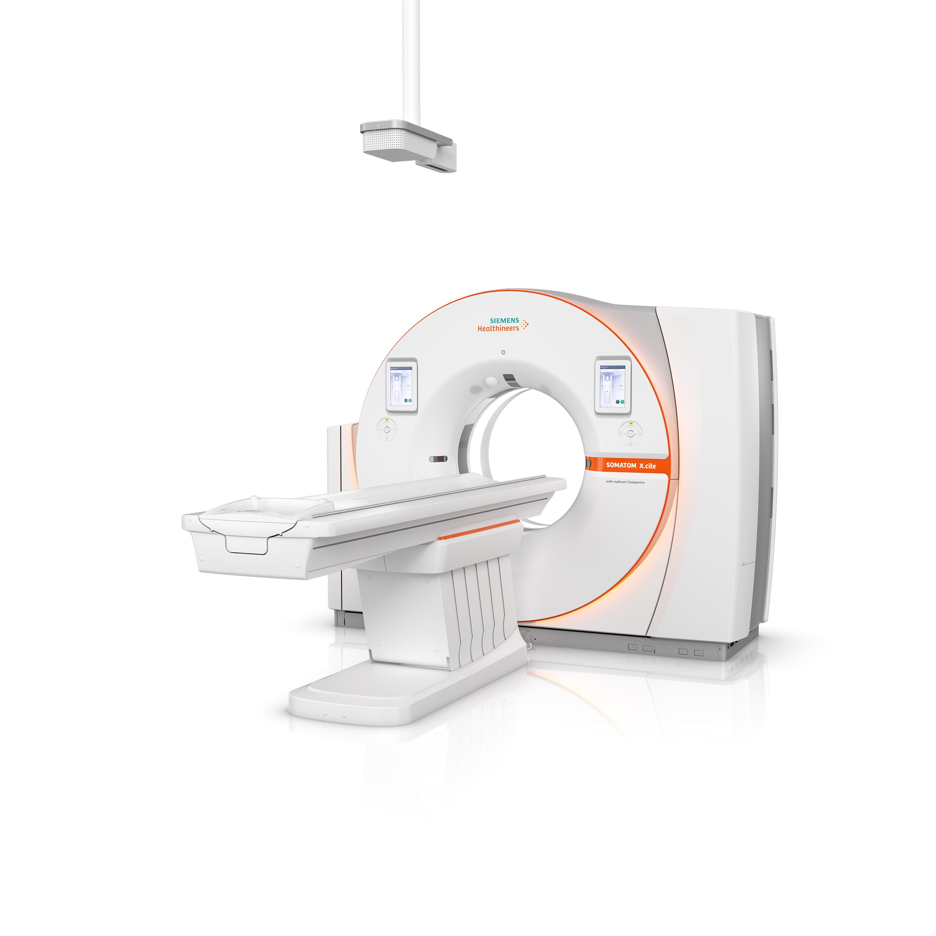
Scanner Features
Philipp Fischer, Head of Computed Tomography, Siemens Healthineers, described the motivation behind the design of Somatom X.cite single-source CT scanner and the myExam Companion; provision of customers with best technology and enabling precision medicine with the highest quality exams and lowest dose.
“It allows us to get deeper and deeper into patient pathology for best diagnostics results and treatment paths,” he said.
myExam Companion, the new intelligent user guidance system lets users utilise the full potential not only of the new Somatom X.cite but also of the Somatom go. scanner platform, already in use today. The system allows sharing of established examination protocols between scanners easily. Workflows and their results are also more standardised, making it possible to reduce deviations at a patient and user level.
Somatom X.cite has a large 82 cm gantry and soothing lighting. It offers a comfortable environment for patients while Medical Technology Radiology Assistants (MTRAs) prepare and perform scans using removable tablets that are attached to the scanner using magnets. This allows them to stay with patients until right before the scan and ensure preparation is full and thorough.
While the scan is in progress, the patients receive audio-visual support and are instructed via both a speaker and a display on how to breathe. If an MTRA has to leave the area, they can still keep their patients in view via a 2D camera integrated into the housing of the gantry encouraging a sense of patient and staff wellbeing.

Follow-up scan for coronary heart disease patient with multiple bypass: Coronary CME CTA spiral scan.
Copyright: Image courtesy of University Hospital Erlanger, Germany
Improved Workflow
The CT scanner gathers data for each specific exam. It does this through automated access to a 3D camera, and patient data such as gender, age, height alongside other important parameters such as heart rate. This feature helps make the workflow more efficient and can also smooth out any difference in experience, which might exist between different MTRAs. This way, the results that are obtained are of high-quality, even in difficult or complicated diagnostic situations. “We lead the technologist with easy and intuitive questions through the examination. A lot of technicalities are done in the background,” said Fischer.

Stroke assessment Flex 4D Spiral.
Copyright: Image courstesy of University Hospital Zurich, Switzerland
Intuitive Use for Radiology Staff
A key roadblock to tech adoption in radiology is that of the perceived expertise needed to operate AI-driven systems. But as far as this new product is concerned, minimal training is needed for staff to adapt to the scanner. Once they understand the system and realise the benefits that it offers, they can focus on the patient and improved quality of care. “It’s like when you get your first smartphone,” said André Hartung. “It may take some time to to adapt to, but then you don’t want to give it up.”
Somatom X.cite single-source CT scanner and myExam Companion work together to gather patient data in the background while radiology staff can focus on the person they have in front of them. The high quality of images supports better and faster diagnostics. “You have to be quick in neurological diseases. Our department was a good testing ground for the scanner,” said Prof. Dr. Christoph Stippich, Director of the Department of Neuroradiology, USZ.

Low kV imaging with high power reserves, low kV even for bigger patients possible.
Copyright: Image courtesy of Clinica Universidad de Navarra, Pamplona, Spain
Patient Benefits
The new Somatom X.cite, featuring the Vectron x-ray tube, and a large gantry, offers high image quality. The 3D camera allows radiologist staff to position the patient properly in the isocenter. “I call it the one-size-fits-all scanner,” explained Prof. Dr. Hatem Alkadhi, Senior Physician Radiology, USZ. “The FAST 3D camera in Somatom X.cite aids in optimally placing the patient in the right position. Presently, around 95% of patients are not positioned properly, and this affects the dose of subsequent CTs. It can increase dose by 30-40% if done manually.”
Somatom
X.cite and myExam Companion have been clinically approved by the FDA and will
be available from December 2019.
Image credit: Siemens Healthineers


