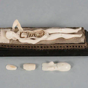
Manikins or small anatomical sculptures, often made of ivory or animal bone, are said to have been used as tools for the study of medical anatomy as far back as four centuries ago. Still, not much is known about the origins of these manikins, which makes them objects of curiosity and highly-prized collector's items.
With advancement in imaging technology, researchers have found a way to better study what these 17th-century manikins are actually made of, without causing damage to the fragile pieces, according to a new study being presented at the annual meeting of the Radiological Society of North America (RSNA).
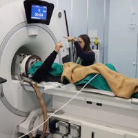
At Duke University, where 22 of 180 known manikins worldwide can be found, Dr Fides R. Schwartz, research fellow in the Department of Radiology and colleagues used micro-CT imaging in order to identify the ivory type used in the Duke manikins and determine whether any repairs or alterations have been made.
Micro-CT has much better resolution compared to standard CT and allows enhanced visualisation of an object's internal features. This technique, according to the researchers, could help them make a more precise estimation of the manikins' age.
"The advantage of micro-CT in the evaluation of these manikins enables us to analyse the microstructure of the material used," Dr Schwartz explained. "Specifically, it allows us to distinguish between 'true' ivory obtained from elephants or mammoths and 'imitation' ivory, such as deer antler or whale bone."
Most of the manikins in the Duke collection were purchased in the 1930s and 1940s by Duke thoracic surgeon Josiah Trent, MD, and his wife Mary Duke Biddle Trent, prior to the 1989 ivory trade ban. Trent's granddaughters donated the manikins to the university, where the sculptures have spent most of their time in archival storage boxes or behind display glass because, as the researchers noted, these items are too fragile for regular handling.
For this study, Dr Schwartz and her team scanned all 22 manikins with micro-CT. They found that 20 out of the 22 manikins were composed of true ivory alone, while one manikin was made entirely of antler bone and another manikin contained both ivory and whale bone components.
There were also metallic components found in four of the manikins, and fibres in two. Moreover, 12 manikins contained hinging mechanisms or internal repairs with ivory pins, and one manikin had a long detachable pin disguised as a hairpiece.
Identifying non-ivory components in the manikins may provide more accessibility to carbon dating, allowing the researchers to more accurately estimate the age of some of the manikins without damage to the fragile pieces.
Additionally, the researchers hope to acquire 3D scans to create digital renderings and enable subsequent 3D printed models.
"Digitising and 3D printing them will give visitors more access and opportunity to interact with the manikins and may also allow investigators to learn more about their history," Dr Schwartz said.
Source: RSNA
Image credit: RSNA
Latest Articles
RSNA 2019 mannikins 3D printing 3D scans imaging
Modern-day imaging provides insights into the materials used in 17th-century anatomical models.


