Introduction
Ultrasound contrast imaging is becoming increasingly important in clinical applications and in researches. Traditional contrast agent
ultrasound imaging technologies, such as pulse inversion/phase inversion
imaging and coded contrast imaging, have focused on the second harmonic
properties of the contrast agents and tissues. Although it has proven that
these technologies are very helpful, it has been recognized that the second
harmonic signals provide limited performance relative to contrast agent
signal strength. The most popular contrast imaging feature CPS requires
system’s good performance on the cancellation of tissue fundamental
signal.However, its spatial resolution is not as good as the 2nd harmonic
contrast imaging.
Although the contrast agent signal can add important information
to the ultrasound image for clinical diagnosis, the ideal process
would be to simultaneously detect the signatures that are unique to contrast
agents and unique to tissue. This would thereby allow the clinician to view the
contrast agent signals separately from the tissue signals.
A new technology, called Ultra-Wideband Nonlinear contrast
imaging (UWN), is now available on Mindray DC8 platform that processes the
combination of the nonlinear fundamental, the 2nd harmonic signals, and the
higher order harmonic signals that are generated by ultrasound contrast
agents. It works very well both at low MI and high MI on various
ultrasound contrast agents. With this new technology Mindray’s DC8 provides
excellent agent-to-tissue specificity of contrast imaging. It achieves the
premium contrast imaging performance by simultaneously processing received
signals from multiple transmitted pulses of varying phase (or polarity) and
amplitude. The precise control of transmit pulse amplitude and phase and
the modulation of the received echoes allow the system to separate
the strong tissue linear fundamental signal from the contrast nonlinear
signals, including nonlinear fundamental and the 2nd harmonic from the contrast
agent. The nonlinear fundamental response shows bubble’s nonlinear behavior of
higher order nonlinearity at 3rd and other higher odd orders, but generates the
new frequency same as the transmitted pulse. By combining the detection of the
nonlinear fundamental signal and the 2nd harmonic signal from
contrast agent we can significantly improve contrast imaging’s Contrast
Tissue Ratio, CTR, and achieve the excellent contrast resolution and
detail resolution.
The uniqueness of the UWN technology is to separate fundamental
tissue signal from the nonlinear contrast agent signals by modulating the received echoes that are transmitted
at different phase and amplitude. The separation is achieved by
proprietary combination and modulation of the returned echoes from multiple
pulses. A low-pass filter can be easily used to filter out the
linear fundamental signal in the modulated echo signals from the contrast
agent.
UWN Technology
The UWN contrast imaging feature on DC8 system transmits
different pulse sequences with the varying pulse amplitude and phase to
support effective tissue signal rejection and bubble signal extraction by
combining and modulating the returned echoes. UWN has designed many
different pulse sequences for different imaging characteristics. A typical example of UWN contrast imaging
transmitted pulse sequence is as follows:
[ a -1 1 1-a ],
where 0 < a < 1.
In the above expression, the maximum transmit magnitude is
normalized to ‘1’, and ‘-‘ represents the opposite polarity, or 1800
opposite phase. Figure 1 shows the transmit waveforms in the transmitted pulse
sequence. The system transmits the pulse sequence in a designed order and receives the returned echoes accordingly. In low MI imaging the main signal component from tissue is linear component. However, the signal from contrast bubble contains both linear and nonlinear components.
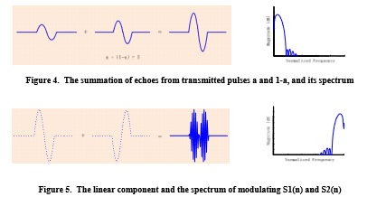
In DC8 the system combines two echo signals from the transmitted pulses with the amplitude of a and 1-a to form a new data set, S1(n). Then, a modulation process is implemented between S1(n) and the returned echo, S2(n), from the transmit pulse of ‘-1’. The modulation process takes the odd time index data from S1(n) and even time index from S2(n) to form a modulated data set. Figure 3 shows the process how to extract the nonlinear signals by such modulation. For the simplicity, the received echo signals are labeled as the echoes from the transmitted pulses [ a -1 1 1-a]. In Figure 3, the upper part is the diagram of how to extract the nonlinear fundamental from contrast agent and the lower part is the diagram of how to extract the 2nd harmonic signal.
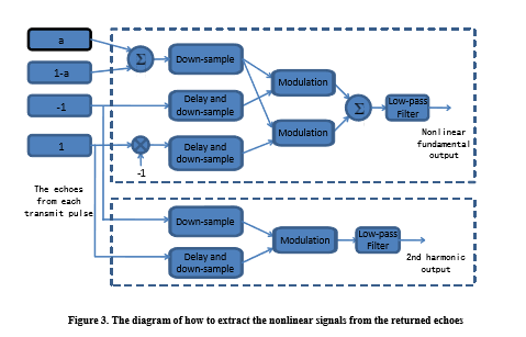
1. Extract the nonlinear fundamental from the echoes of contrast agent
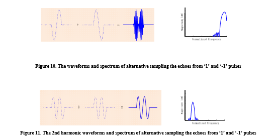
First, to form a full amplitude linear signal, S1(n), by adding the echoes from two transmitted pulses at the amplitude of a and 1-a. The amplitude of the linear component in S1(n) is ‘1’, and the nonlinear fundamental generated by the 3rd order nonlinearity in this new formed signal is proportional to a3+(1‐a)3, where 0 < a < 1. Then, alternatively sample the data between S1(n) with time index 2*i-1, where i = 1, 2, 3, … N, and S2(n), the echo signal from the transmit pulse ‘-1’ at the time index 2*i, where i = 1, 2, 3, … N. After this process, the linear component will be shifted away from its original position in the spectrum. However, the asymmetric part of nonlinear fundamental will stay at its original position in the spectrum; see the plots from Figure 3 to Figure 8.
1). Modulating the linear component
It is shown in Figure 5 the linear component is modulated away from its original position in the spectrum.
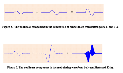
2). Modulating the non-linear component
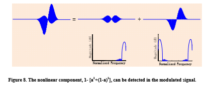
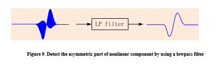
In Figure 8, the modulated nonlinear component can be divided into two parts. One is the symmetric part of [a3+(1‐a)3] and -[a3+(1‐a)3], which is shown in the center plot. The symmetric part of nonlinear component is modulated away from its original position in the spectrum. The other part is the asymmetric part of 1- [a3+(1‐a)3], which stay at the original position in the spectrum. By simply using a low pass filter, the asymmetric part of nonlinear component can be detected, see Figure 9 below.
3). Double NLF sensitivity by adding another NLF detection
Mindray UWN pulse sequence doubles the NLF sensitivity with a well designed transmitted pulse sequence and the receive modulation process. A similar NLF detection process can be done to combine the echo from the 3rd transmit pulse at the full amplitude of ‘1’ by multiplying with a negative sign ‘-‘. The new resulted NLF signal will be combined with the result in the similar process described previously. By adding two NLF detections it can double the NLF sensitivity..
2. Extract the 2nd harmonic contrast agent signal from the echoes
By alternatively sampling the odd time index and even time index between two echoes from the full amplitude transmitted pulses of ‘1’ and ‘-1’, the linear fundamental is modulated away from its original position in the spectrum. However, the 2nd harmonic signal stays at the original position in the spectrum, see the plots below.
Since the 2nd harmonic signals from the transmitted pulses of ‘1’ and ‘-1’ are in phase, they are not modulated.
The 2nd harmonic stays at the same position in the spectrum after the modulation. A low-pass filter can be inserted into the signal path to filter out the modulated fundamental signal and detect the 2nd harmonic.
3. Frequency synthesis of nonlinear contrast agent signals
Two approaches can be used to combine the nonlinear fundamental signal and 2nd harmonic signal from contrast agent. One is the so called frequency compounding, in which after the detection of the nonlinear fundamental and the 2nd harmonic two signals are down-converted to base band and combined after the amplitude detection. The other is to do wideband detection, in which each component is down-converted and form a wideband nonlinear contrast imaging in base band.
In summary, by utilizing this pulse sequence strategy of modulation, the UWN technology can effectively separate tissue fundamental signal from contrast agent nonlinear signals. On DC8 system UWN technology provides the unique detection approach to detect both nonlinear fundamental and the 2nd harmonic separately and combines them in a distinguished way. Comparing to other contrast imaging technologies, UWN has the following advantages:
- The nonlinear fundamental signal generated by contrast agent’s 3rd order nonlinearity is the strongest nonlinear signal from bubbles and falls into transducer’s receive band. UWN fully uses of its transducer’s bandwidth to receive full bandwidth of both nonlinear fundamental and second harmonic signals. By combining them together using different strategies system can provide the best spatial resolution and CTR for the needs.
- The nonlinear fundamental signal is generated uniquely by the contrast agent, not from tissue. By modulating the received echoes, UWN can completely separate the tissue linear fundamental from the nonlinear fundamental and the 2nd harmonic signals generated by the contrast agent. As the consequence, system can display linear fundamental signal; the nonlinear fundamental; and 2nd harmonic separately.
- UWN is an attractive technology to be used for both low MI and high MI applications, as well as for high frequency imaging, where the useful harmonic frequencies would be beyond the bandwidth of today’s state-of-the art transducer. High frequency imaging offers spatial resolution on the order of several hundred microns – which may be applied to areas such as the human breast, thyroid, testicle, or carotid arteries, etc. The emerging field of small animal imaging for use in genomics, pharmaceutical, or other research studies can also benefit from improved resolution at high frequencies with excellent agent-to-tissue specificity.
Examples of Clinical Result
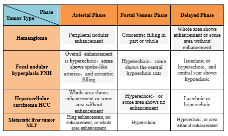
UWN contrast imaging on DC8 in liver application provides good clinical result for tumor classification and diagnosis. The following is a list of common tumors on liver and the blood supplies behaviors at different phase of contrast imaging in liver application:
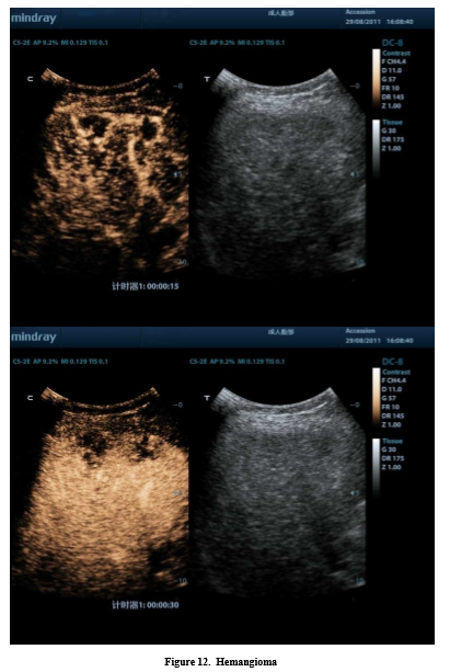
Hemangioma
Hemangioma is the most popular benign tumor on liver. Conventional ultrasound image shows hyperechoic on most of hemangioma (67%-79%). In Contrast imaging at arterial phase it shows fast or slow peripheral nodular enhancement or facelift. Gradually with time it becomes ‘nodular’ or ‘cotton wool’ looking and extends toward the internal of the lesion, shown in Figure 12.
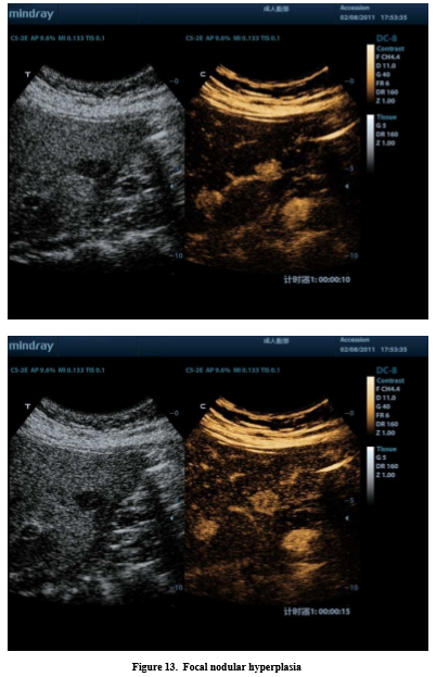
Focal Nodular Hyperplasia
Focal Nodular Hyperplasia is constituted by the proliferation of epithelial cells in benign. Conventional ultrasound imaging shows no abnormal echo from the inside. Compared with the surrounding liver parenchyma it shows isoechoic, or slightly lower or higher echoic, and the boundary is not very clear. In grey-level ultrasound imaging it is not easy to find the lesion. However, contrast imaging can show the special characteristics on Focal Nodular Hyperplasia. In arterial phase the lesion is enhanced rapidly from the center and spread to the surrounding area and fill the surrounding area uniformly to make it more hyperechoic, shown in Figure 13. Typically, it shows ‘radiation’ shape, or ‘stellate’ shape of the internal vessels.
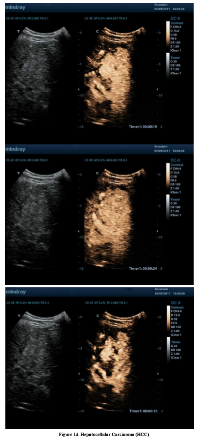
Hepatocellular Carcinoma (HCC)
Hepatocellular Carcinoma, mostly occurres on the persons who have chronic hepatitis, or cirrhosis. It has various appearances in conventional ultrasound imaging and is difficult to be distinguished from the other liver tumors. In contrast imaging it shows uniform hyperechoic whole enhancement at arterial phase, see Figure 14.
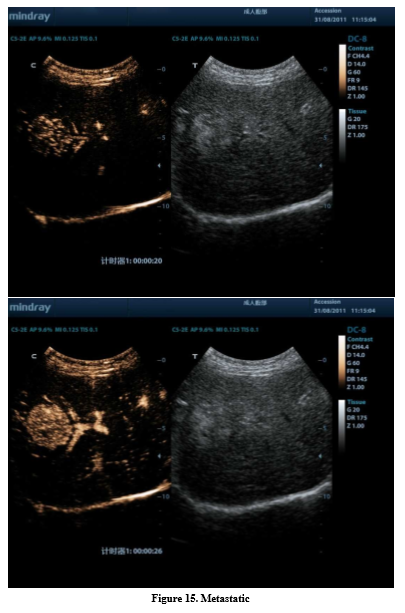
Metastatic
Metastatic is malignant tumor of the secondary of the liver, or spread to the liver from the other organs. The most common way is the intestinal tumor spreads to the liver by the portal blood flow. Contrast imaging shows some special characteristics on Metastatic. The lesion is often rapidly ring enhanced or whole area enhanced at arterial phase. Enhancement peak often shows a hyperechoic ring shape or overall hyperechoic. As far as the duration time of the enhancement, Metastatic shows fast wash in and out. Compared to HCC, Metastatic’s wash in and out is faster than HCC. Normally, the subside starts at about 25 second after the injection of the contrast agent, shown in Figure 15.