Remarkable image quality, right at the point of care.
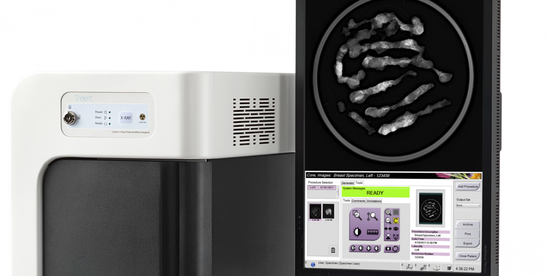
The Trident system revolutionizes breast tissue imaging by incorporating a micro-focused tube, unique specimen image processing algorithms and amorphous selenium direct digital detector. The result is sharp, high quality images for rapid specimen verification.
The Trident system features an intuitive user interface ideal for non-technical operators, with one-touch X-ray automatic exposure control (AEC), a lighted specimen X-ray chamber and large active imaging area. The system incorporates easy-to-use software with a simple, yet robust toolset.
Designed to maximize workflow efficiencies, the Trident system enables quick communication and decision-making; with the push of one button, you can export images to the Hologic SecurView® DX diagnostic workstation, PACS and other preferred output devices, or to a printer for hard-copy review.
Superb image quality for rapid verification with maximum confidence
- One-touch X-ray control with AEC for fast image acquisition.
- Large, 12 x 14 cm active imaging area; accommodates a wide range of surgical specimens and core biopsy samples.
- Innovative enhanced visualization tool with five levels of image optimization for added sharpness and lesion conspicuity.
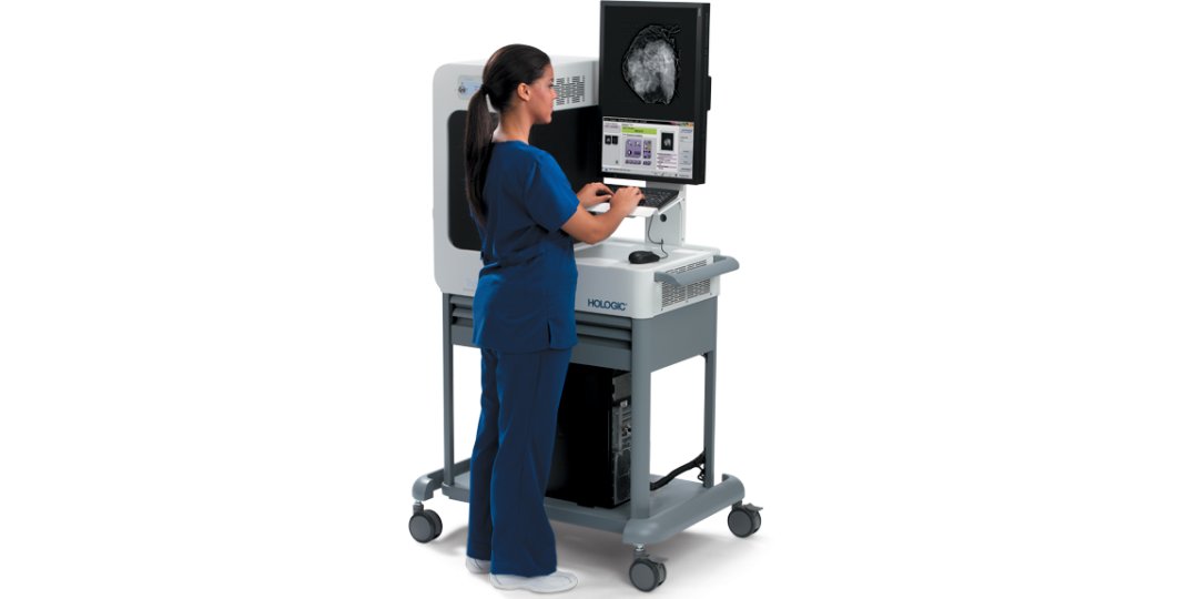
- Fully integrated, maneuverable and ergonomic workstation.
- User-friendly operator interface with minimal procedure steps to streamline workflow.
- Ideal for radiologists, breast surgeons and pathologists.x


")


