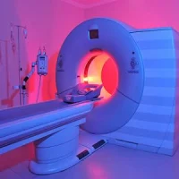
In a pioneering study, researchers in Japan have found a way to use x-rays in examining soft tissues – not just dense materials like bones – similar to how ultrasound or magnetic resonance imaging (MRI) work.
You might also like: AI+Radiologist: Improved Screening Interpretation
Both ultrasound and MRI make use of elastography, a noninvasive imaging approach for investigating the stiffness and elasticity of soft tissue, enabling doctors to identify lesions and other deep tissue problems.
While x-ray elastography may be possible "in principle", as shown in previous studies, the Japanese researchers' work is the first ever to apply the concept for real-world visualisation of the stiffness of soft tissue, according to an article published in the journal Applied Physics Express.
Of note, compared to ultrasound, x-rays provide much greater resolution – on the order of tens of micrometres (millionths of a metre) instead of millimetres (mere thousandths of a metre), said the Japanese research team.
"This greater precision doesn't just mean identification of much smaller or deeper lesions, but, importantly for patients, because smaller lesions can be newer ones, potentially also much earlier on in a disease or condition," explained lead researcher Wataru Yashiro, an associate professor from the Institute of Multidisciplinary Research for Advanced Materials (IMRAM) of Tohoku University.
The researchers said their technique, called dynamic x-ray elastography, relies on "shear wave" propagation. Shear waves are similar to those that occur when you whip a rope up and down quickly. Shear waves travel faster through stiffer tissue than through softer tissue.
Cancerous tumours, lesions from cirrhosis of the liver and hardened arteries are stiffer than the surrounding healthy tissue, according to the researchers, thus identifying where the waves pass through tissue more slowly, clinicians are able to pinpoint these stiffer tissues.
Yashiro and colleagues are eyeing to further develop their technique to produce 3D visualisations, and then move towards the manufacture of x-ray elastography medical diagnostic equipment.
Source: Tohoku University
Image credit: iStock
ReferenceL Kamezawa
C, Yashiro W et al. (2020) X-ray elastography by visualizing propagating shear
waves. Appl. Phys. Express 13 042004. DOI
https://doi.org/10.35848/1882-0786/ab7e06