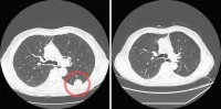
A recent study has confirmed the effectiveness of x-ray phase tomography with synchrotron radiation in detecting cancerous tissue in a mouse model. "Phase-contrast imaging" produced images with much enhanced contrast, making a clear differentiation between cancerous tissue and healthy tissue, according to researchers. The findings have been reported in the Journal of Applied Physics.
X-ray phase tomography delivers images with high contrast compared to conventional CT (computed tomography) scan images. In the study, the researchers compared three different x-ray phase tomography methods: x-ray grating interferometry, propagation-based phase tomography with single-distance phase reconstruction, and holotomography. The investigators put these three techniques to the test by examining cancerous tissue from a mouse model and an entire rat's heart.
The research team wanted to determine which technique performs best in terms of spatial resolution and visualisation/quantification of relevant features in the samples. They also examined other factors such as the simplicity of the setup, and how the data are acquired and analysed with each method.
For their study, the investigators opted to exploit synchrotron radiation, which produces higher-quality x-rays than conventional x-ray generators, such as those used in hospitals. Synchrotron radiation can be likened to the monochromatic, collimated and intense light produced by lasers, while conventional x-ray generators are more analogous to common light bulbs used in most homes, said lead author Irene Zanette, a scientist affiliated with the European Synchrotron Radiation Facility (ESRF) in France and the Technische Universität München (TUM) in Germany.
Although the study was performed using synchrotron radiation, Zanette noted that the same techniques are compatible with both polychromatic and divergent beams and can also be implemented at conventional x-ray sources. The research team used the phase-contrast x-ray imaging technique, which works by making the x-ray beam interfere while it propagates from sample to detector. "This interference is fundamental because it encodes precious information on the phase of the X-ray waves," Zanette explained.
In contrast, conventional x-ray imaging — of the sort performed at hospitals and airports — doesn't use phase information. Instead, it relies only on the attenuation of the amplitude (reduction in intensity) of the x-ray waves by the object under study to generate image contrast. As Zanette pointed out: "More detailed information is contained in the phase than the amplitude, so it enables us to obtain images with much greater contrast and clearly differentiates cancerous tissue from healthy tissue."
Zanette's team reported these key findings:
The team therefore regards the three phase tomography methods they tested as being complementary. What is important is to be able to choose the technique that suits "your specific purposes," according to co-author Bert Müller, group leader of the Biomaterials Science Centre in Switzerland.
"Our research should help provide guidance for other researchers in developing an ideal phase-contrast imaging method, which will be adopted by hospitals in the future," noted Zanette, currently a postdoctoral scientist in biomedical physics at TUM in Germany.
Source: AlphaGalileo.org
Image Credit: Irene Zanette/Technische Universität München
X-ray phase tomography delivers images with high contrast compared to conventional CT (computed tomography) scan images. In the study, the researchers compared three different x-ray phase tomography methods: x-ray grating interferometry, propagation-based phase tomography with single-distance phase reconstruction, and holotomography. The investigators put these three techniques to the test by examining cancerous tissue from a mouse model and an entire rat's heart.
The research team wanted to determine which technique performs best in terms of spatial resolution and visualisation/quantification of relevant features in the samples. They also examined other factors such as the simplicity of the setup, and how the data are acquired and analysed with each method.
For their study, the investigators opted to exploit synchrotron radiation, which produces higher-quality x-rays than conventional x-ray generators, such as those used in hospitals. Synchrotron radiation can be likened to the monochromatic, collimated and intense light produced by lasers, while conventional x-ray generators are more analogous to common light bulbs used in most homes, said lead author Irene Zanette, a scientist affiliated with the European Synchrotron Radiation Facility (ESRF) in France and the Technische Universität München (TUM) in Germany.
Although the study was performed using synchrotron radiation, Zanette noted that the same techniques are compatible with both polychromatic and divergent beams and can also be implemented at conventional x-ray sources. The research team used the phase-contrast x-ray imaging technique, which works by making the x-ray beam interfere while it propagates from sample to detector. "This interference is fundamental because it encodes precious information on the phase of the X-ray waves," Zanette explained.
In contrast, conventional x-ray imaging — of the sort performed at hospitals and airports — doesn't use phase information. Instead, it relies only on the attenuation of the amplitude (reduction in intensity) of the x-ray waves by the object under study to generate image contrast. As Zanette pointed out: "More detailed information is contained in the phase than the amplitude, so it enables us to obtain images with much greater contrast and clearly differentiates cancerous tissue from healthy tissue."
Zanette's team reported these key findings:
- For each specimen, the spatial resolution derived from the characteristic morphological features is about twice as good for holotomography and single-distance phase reconstruction compared to x-ray grating interferometry.
- X-ray grating interferometry data generally provide much better contrast-to-noise ratios for anatomical features, excel in fidelity of the density measurements, and are more robust against low-frequency artifacts than holotomography.
The team therefore regards the three phase tomography methods they tested as being complementary. What is important is to be able to choose the technique that suits "your specific purposes," according to co-author Bert Müller, group leader of the Biomaterials Science Centre in Switzerland.
"Our research should help provide guidance for other researchers in developing an ideal phase-contrast imaging method, which will be adopted by hospitals in the future," noted Zanette, currently a postdoctoral scientist in biomedical physics at TUM in Germany.
Source: AlphaGalileo.org
Image Credit: Irene Zanette/Technische Universität München
References:
Lang S, Zanette, I Dominietto M et al. (2014) Experimental comparison of grating- and propagation-based hard X-ray phase tomography of soft tissue. J Appl Phys 116(15). http://dx.doi.org/10.1063/1.4897225
Latest Articles
X-ray, Radiation, CT scans, synchrotron, tomography
A recent study has confirmed the effectiveness of x-ray phase tomography with synchrotron radiation in detecting cancerous tissue in a mouse model. "Phase-...