


Modern imaging techniques (CT and MRI) have enabled researchers to investigate hearts more than 400 years old found at an archaeological site, helping them learn about the health conditions of the people buried there. Of the five hearts examined, one heart appeared healthy and showed no signs of disease. Three of the hearts did show signs of disease, as plaque was found on the coronary arteries. The fifth heart had been poorly preserved and, therefore, could not be studied.
The research, to be presented Wednesday, 2 December, at the annual meeting of the Radiological Society of North America (RSNA), is the first to use radiological examination to study archaeological hearts.
Five heart-shaped lead urns were found by archaeologists in grave sites in France that date back to the late 16th or early 17th century. Inside each urn was a preserved human heart. A team of radiologists, including one with a background in forensics, was called in to examine the hearts. Additional researchers, including forensic physicians, archaeologists, pathologic physicians and physicists, were brought in from the Molecular Anthropology and Synthesis Imaging and the Institute of Metabolic and Cardiovascular Diseases.
The research team used MRI and CT to obtain clinical images of the hearts. While the images were impressive, due to the embalming materials used to preserve the hearts, very little health information could be obtained. The team then "cleaned" the hearts to remove the embalming material. MRI and CT scans were redone.
On the new set of CT images, researchers were able to identify the different heart structures, such as chambers, valves and coronary arteries. Once the tissue was rehydrated, researchers were better able to identify myocardial muscles with MRI. Classic techniques, such as dissection, external study and histology, were also used to examine the heart tissues.
"Since four of the five hearts were very well preserved, we were able to see signs of present-day heart conditions, such as plaque and atherosclerosis," said study author Fatima-Zohra Mokrane, MD, radiologist at Rangueil Hospital at the University Hospital of Toulouse in France.
When and Where
Dr. Mokrane will present "Is it Possible to Investigate Archeological Hearts Using CT and MRI? About Five Archeological Hearts" during the Cardiac (General Topics) session on Wednesday 2 December, 11.30-11.40am S504AB, South Building Level 5.
Iphigenia Papaioanou
Editorial Project Director, HealthManagement.org
Source: RSNA
Image credit (top): Rozenn Colleter, PhD/INRAP
The research, to be presented Wednesday, 2 December, at the annual meeting of the Radiological Society of North America (RSNA), is the first to use radiological examination to study archaeological hearts.
Five heart-shaped lead urns were found by archaeologists in grave sites in France that date back to the late 16th or early 17th century. Inside each urn was a preserved human heart. A team of radiologists, including one with a background in forensics, was called in to examine the hearts. Additional researchers, including forensic physicians, archaeologists, pathologic physicians and physicists, were brought in from the Molecular Anthropology and Synthesis Imaging and the Institute of Metabolic and Cardiovascular Diseases.
The research team used MRI and CT to obtain clinical images of the hearts. While the images were impressive, due to the embalming materials used to preserve the hearts, very little health information could be obtained. The team then "cleaned" the hearts to remove the embalming material. MRI and CT scans were redone.
On the new set of CT images, researchers were able to identify the different heart structures, such as chambers, valves and coronary arteries. Once the tissue was rehydrated, researchers were better able to identify myocardial muscles with MRI. Classic techniques, such as dissection, external study and histology, were also used to examine the heart tissues.
"Since four of the five hearts were very well preserved, we were able to see signs of present-day heart conditions, such as plaque and atherosclerosis," said study author Fatima-Zohra Mokrane, MD, radiologist at Rangueil Hospital at the University Hospital of Toulouse in France.
When and Where
Dr. Mokrane will present "Is it Possible to Investigate Archeological Hearts Using CT and MRI? About Five Archeological Hearts" during the Cardiac (General Topics) session on Wednesday 2 December, 11.30-11.40am S504AB, South Building Level 5.
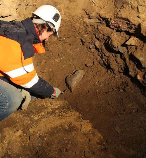
Archaeologist, Dr. Rozenn Colleter, excavating the fifth heart-shaped lead urn. Image credit: Gaétan LeCloire/INRAP
Iphigenia Papaioanou
Editorial Project Director, HealthManagement.org
Source: RSNA
Image credit (top): Rozenn Colleter, PhD/INRAP
Latest Articles
healthmanagement, CT, MRI, heart disease, RSNA 2015, #RSNA15, archaeology
Modern imaging techniques (CT and MRI) have enabled researchers to investigate hearts more than 400 years old found at an archaeological site, helping them learn about the health conditions of the people buried there. Of the five hearts examined, one hear


