Magnetic resonance imaging (MRI) is a noninvasive way to measure iron levels in the brains of people with attention deficit hyperactivity disorder (ADHD), according to a study being presented today at the annual meeting of the Radiological Society of North America (RSNA). Researchers said the method could help physicians and parents make better informed decisions about medication.
ADHD is a common disorder in children and adolescents that can continue into adulthood. Symptoms include hyperactivity and difficulty staying focused, paying attention and controlling behavior. The American Psychiatric Association reports that ADHD affects 3 to 7 percent of school-age children.
Psychostimulant medications such as Ritalin are among the treatments used. Psychostimulants affect levels of dopamine, a neurotransmitter in the brain associated with addiction.
"Studies show that psychostimulant drugs increase dopamine levels and help the kids that we suspect have lower dopamine levels," said Vitria Adisetiyo, Ph.D., postdoctoral research fellow at the Medical University of South Carolina. "As brain iron is required for dopamine synthesis, assessment of iron levels with MRI may provide a noninvasive, indirect measure of dopamine."
Dr. Adisetiyo and colleagues measured brain iron in 22 children and adolescents with ADHD and in a control group 27 healthy children and adolescents using an MRI technique called magnetic field correlation (MFC) imaging. The technique is relatively new, having been introduced in 2006 by study co-authors and faculty members Joseph A. Helpern, Ph.D., and Jens H. Jensen, Ph.D.
"MRI relaxation rates are the more conventional way to measure brain iron, but they are not very specific," Dr. Adisetiyo said. "We added MFC because it offers more refined specificity."
The results showed that the 12 ADHD patients who had never been on medication had significantly lower MFC than the 10 ADHD patients who had been on psychostimulant medication or the 27 typically developing children and adolescents in the control group. In contrast, no significant group differences were detected using relaxation rates or serum measures. The lower brain iron levels in the non-medicated group appeared to normalize with psychostimulant medication.
MFC imaging's ability to noninvasively detect the low iron levels may help improve ADHD diagnosis and guide optimal treatment. Noninvasive methods are particularly important in a pediatric population, Dr. Adisetiyo noted.
"This method enables us to exploit inherent biomarkers in the body and indirectly measure dopamine levels without needing any contrast agent," she said.
If the results can be replicated in larger studies, then MFC might have a future role in determining which patients would benefit from psychostimulants—an important consideration because the drugs can become addictive in some patients and lead to abuse of other psychostimulant drugs like cocaine.
"It would be beneficial, when the psychiatrist is less confident of a diagnosis, if you could put a patient in a scanner for 15 minutes and confirm that brain iron is low," she said. "And we could possibly identify kids with normal iron levels who could potentially become addicts."
Along with replicating the results in a larger population of patients, the researchers hope to expand their studies to look at the relationship between cocaine addiction and brain iron.
Other co-authors are F. Xavier Castellanos, MD, Adriana Di Martino, MD, Kevin M. Gray, MD, Els Fieremans, Ph.D., Ali Tabesh, Ph.D., and Rachael L. Deardorff, MS.
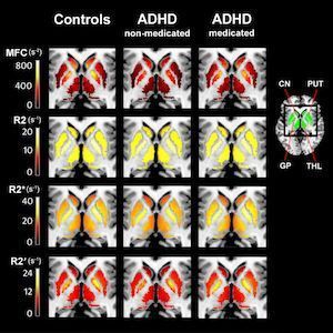
Figure 1: Medication-naïve ADHD patients (ADHD-non-medicated) have reduced striatal (putamen (PUT), caudate nucleus (CN)) and thalamic (THL) magnetic field correlation (MFC) measures of brain iron compared to controls and psychostimulant medicated ADHD patients (ADHD-medicated); Brain iron measures in PUT, CN, THL or globus pallidus (GP) did not differ between controls and psychostimulant medicated ADHD patients. These statistical significant differences are visible in the MFC group maps (top row) but not in conventional relaxation rate maps: R2 (second row), R2*(third row), and R2' (bottom row).
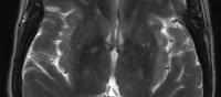
Image credit - top: Siemens Magnetom Trio MRI Scanner
Source: Radiological Society of North America
Latest Articles
MRI, ADHD
Magnetic resonance imaging (MRI) is a noninvasive way to measure iron levels in the brains of people with attention deficit hyperactivity disorder (ADHD),...