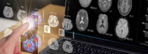
Scientists from Guys’ and St Thomas’, King’s College, London, Imperial College, London and the University of Oxford will use a Philips state-of-the-art MRI scanner combined with specially developed imaging techniques to investigate the wombs of pregnant women and image their babies’ brains, even while the foetuses are moving. To deliver this ambitious project new MRI facilities have been installed at St Thomas’ Hospital, creating a dedicated imaging suite, integrated with the neonatal critical care unit and a wide bore scanner for foetal imaging. Philips has supplied an Achieva TX MRI system and an Avalon foetal monitor, and will be providing ongoing support.
The researchers will use the advanced imaging techniques to map how the human brain grows and forms connections, with the aim of mapping out for the first time how the brain assembles itself, and also observe how brain connections and patterns of activity, seen on older subjects, emerge as babies grow in the womb and just after birth. Up to 1,500 subjects will be studied to examine both normal development, and also explore early signs of disabilities such as autism and attention deficit disorders. The objective is to create a “connectome”, a kind of wiring diagram of the foetal and infant brain showing the formation of brain structures such as the cerebral cortex, where ‘thinking’ occurs, or the hippocampus, which is central to memory, and the connections between them.
Professor David Edwards, director of the Centre for the Developing Brain at King’s said “We want to create a series of atlases showing the human brain at different stages in development. Eventually, we hope to have enough data to compare the brains of children who go on to develop normally with those who develop conditions like autism and attention deficit disorders, so that we can see if such conditions have their origins in the way the brain developed in the womb.”
MRI is already used to image brains of unborn babies, often after ultrasound exams have shown potential abnormalities, and it is also increasingly used to help in the management of premature babies and other vulnerable infants. The information provided is becoming more and more important as new treatments are developed that can improve the outcome for babies at risk of long-term brain damage occurring around the time of birth. However, while established imaging techniques provide detailed anatomical information and reveal if there is structural brain damage, mapping of brain connectivity and emerging brain function is a task at the limits of current capabilities. The team will, therefore, have to develop both advanced imaging methods and new ways to analyse the images to extract the vital connectome information.
Jo Hajnal, professor of imaging science at King’s, a physicist who specializes in imaging subjects who cannot remain still while they are scanned said “We will scan about 1,500 children; about 500 will be foetuses, another 500 will be newborn normal babies, and the rest will be at-risk babies, including 200 premature babies and about 300 babies identified as being at higher risk of development autism.”
Professor Hajnal added, “The foetal scans let us look back into the womb and see how the brain assembles itself, while the normal babies will give us a sense of just what a brain looks like, and how it works when a person enters the world. We can compare these images with what we see in the brains of at-risk babies and maybe find out what makes them different.”
Once the images have been collected, advanced computational analysis methods will be developed and applied to extract connectome information. This critical part of the project will be led by Professor Daniel Rueckert, Department of Computing, Imperial College London, and Professor Steve Smith of Oxford University’s centre for Functional MRI of the Brain.
The developing human connectome project is funded by the European Research Council under the new Synergy Grant Scheme. The Oxford team is also involved in a sister project aimed at mapping the adult human connectome, which is being led from the United States.
Latest Articles
MRI, Philips, Infants
Scientists from Guys’ and St Thomas’, King’s College, London, Imperial College, London and the University of Oxford will use a Philips state-of-the-a...

