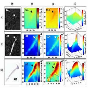
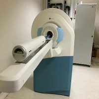
A new study presented at the 2017 Annual Meeting of the Society of Nuclear Medicine and Molecular Imaging (SNMMI) shows a hybrid molecular imaging system uniting three imaging modalities to map the composition of dangerous arterial plaques before they rupture and induce a major cardiac event.
The imaging system has been developed by Stanford University researchers and it unites opitcal, radioluminescence and photoacoustic imaging to evaluate thin-cap fibro atheroma (TCFA). Some plaques associated with atherosclerosis can be quite unstable and if left untreated, can lead to rupture and death. TCFAs' are especially prone to rupture.
Raiyan T. Zaman, PhD, instructor of cardiovascular medicine at Stanford University School of Medicine in Stanford, California, explains that this new system is the first of its kind to be able to detect vulnerable plaque in their earliest stages despite their size, complex biochemistry and morphology. The use of this novel system could change the way coronary artery disease is diagnosed and assessed.
Called the Cirumferential-Intravascular-Radioluminescence-Photoacoustic-Imaging (CIRPI) system, it allows high-acuity optical imaging via beta-sensitive probe and radioluminescent marking to determine inflammation. Photoacoustic imaging provides information about the biological makeup of the plaques.
Study researchers focused on atherosclerotic samples of both human and mouse carotid arteries. They performed CIRPI following injection of fluorine-18 fluorodeoxyglucose (18F-FDG). Plaque composition was delineated through photoacoustic lasers. The results was a never-before-seen 360-degrees perspective of arterial plaque burden.
"This is an important and potentially life-saving tool that could one day be used by interventional cardiologists to identify the appropriate treatment plan for patients at risk of future TCFA rupture," explains Zaman.
Source: Society of Nuclear Medicine
Image Credit: Stanford University School of Medicine and Stanford University Department of Electrical Engineering
Latest Articles
PET, atherosclerosis, hybrid molecular imaging system, arterial plaques
A new study presented at the 2017 Annual Meeting of the Society of Nuclear Medicine and Molecular Imaging (SNMMI) shows that a hybrid molecular imaging system uniting three imaging modalities to map the composition of dangerous arterial plaques before the


