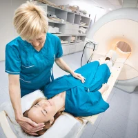
Newer targeted PET tracers have been developed that target disease-specific biomarkers, and allow accurate and sensitive detection of disease. Combined with the superior capabilities of MR imaging to evaluate soft tissue, precision imaging with PET/MRI can change the diagnosis and treatment, says an article in press in the journal PET Clinics.
"With current and continued development in both PET and MR technology, PET/MR imaging will be a large contributor in precision medicine now and in the future," the article points out.
Precision medicine is loosely defined as an approach to disease treatment and prevention that takes into account individual variability in the patient’s tumour. As therapies have become more disease specific, diagnostic imaging must continue to advance with tailored disease-specific agents.
Early studies have shown combining the use of these targeted precision medicine PET agents with MR imaging has some definite advantages compared with PET/computed tomography (CT). Unlike the low intrinsic soft tissue contrast of CT, the superior soft tissue contrast of MRI allows for the improved assessment of fine anatomic detail and can provide additional information on tissue composition and function not possible with CT, the article explains.
"In allowing for more precise evaluation of disease-specific markers in combination with excellent anatomic detail, PET/MR imaging offers a way not only to identify those patients who may benefit from targeted treatment but also a better method of evaluating disease response," the report says.
In addition to the anatomic detail and contrast enhancement, MRI is capable of providing functional information with diffusion-weighted imaging (DWI), perfusion imaging, and spectroscopy not possible with CT and without the use of ionising radiation. DWI has proven particularly valuable in the assessment of lesion cellularity, and may be used as a whole-body screening technique for the detection of neoplastic lesions, including small lesions less than 10 mm in diameter. Magnetic resonance spectroscopy’s ability to detect metabolites can also help to differentiate normal versus abnormal tissue.
Currently, PET/MRI is primarily used with less specific nontargeted molecular imaging agents. Through the imaging of cell processes, nontargeted molecular imaging can be used to identify abnormal cell physiology and monitor cellular changes over time. The most common cellular processes imaged by nontargeted molecular imaging and PET/MRI are glucose metabolism, lipogenesis, cellular proliferation or DNA synthesis, and cellular hypoxia. The most commonly used nontargeted agent is fluorodeoxyglucose (FDG) PET.
Although clinical studies have suggested that targeted molecular imaging can provide a useful method for monitoring targeted therapy in PET/MRI, there are currently several barriers limiting the research and clinical use of PET/MRI in precision medicine, the article notes. Major barriers include the relatively small numbers of PET agents approved by the U.S. Food and Drug Administration, accessibility to PET/MRI scanners, and relative lack of data supporting the use of PET/MRI compared with PET/CT using these tracers.
Still, progress is being made in demonstrating the superiority of PET/MRI compared with PET/CT, including applications in targeted evaluation of prostate cancer, neuroendocrine tumours (NETs), and parathyroid disease. "It is hoped that these studies and larger clinical trials will lay the groundwork for determining clinical indications and standardising PET/MR imaging protocols," according to the article.
Source: PET Clinics
Image Credit: MR Solutions
References:
Huo, Eugene et al. (2017) The Role of PET/MR Imaging in Precision Medicine. PET Clinics. doi.org/10.1016/j.cpet.2017.05.006
Latest Articles
PET, MRI, Precision Imaging
Newer targeted PET tracers have been developed that target disease-specific biomarkers, and allow accurate and sensitive detection of disease. Combined with the superior capabilities of MR imaging to evaluate soft tissue, precision imaging with PET/MRI ca