Executive Summary
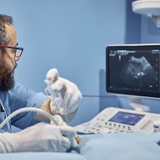
Proceduralultrasound, according to Dr. Nikhani, not only helps clinicians identify the relevant anatomy and pathology before proceeding with invasiveprocedures, but it also aids accurate execution through direct visualisation as the needle advances towards the target vessel. "Quite simply, the effect is similar to turning on a car’s headlights at night to navigate safely to the desired destination," Dr. Nikhani writes. The practice is also recommended by the American College of Emergency Physicians (ACEP) as this enables a “one stick standard” for faster, safervascularaccess to accelerate patientcare.
You may also like: Bedside Ultrasonography: 6 Success Factors for Implementation
In 2016, ACEP issued a policy statement highlighting that the benefits of proceduralultrasound, performed at the bedside, include “improved patient safety, decreased procedural attempts and decreased time to perform many procedures in patients whom the technique would otherwise be difficult."
Achieving rapid vascular access is particularly critical for providing optimal care for critically and unstable patients. Newer techniques now allow subclavian catheters to be placed under ultrasound guidance, though internal jugular vein access is the most commonly performed due to its ease. Of note, failure rates of emergent peripheral intravenous (PIV) access of 10-40% have been reported in the literature.
"However, ultrasound-guided PIV, which is a standard practice at the hospital where I work," the author says, "can help such patients avoid unnecessary CVCs and their associated risks." Furthermore, findings from a randomised trial of emergency department patients with difficult vascular access and other studies offer a powerful argument for widespread adoption of ultrasound-guided PIV as an evidence-based safety practice to reducecosts and accelerate care for patients who need it the most. "And if you or a loved one ever needed a central or peripheral line for emergency treatment, wouldn’t you want ultrasound at the bedside and a medical provider who was firmly committed to achieving the one-stick standard?" said Dr. Nikhani.
Source: Full article will be published 22 February 2019 in HealthManagement.org
Image credit: iStock
References:
Latest Articles
Telehealth, Ultrasound, Critical Care, patient safety, patient care, CVC, vascular access, ACEP, invasive procedures, American College of Emergency Physicians, Ultrasound-guided CVC Placement, procedural ultrasound, catheterisations, Banner Telehealth Services, Los Angeles, ultrasound visualisation, one stick standard, rapid vascular access, subclavian catheter, PIV, ultrasound-guided PIV, David Geffen School of Medicine
The majority of critical care patients (80%) undergo central venous catheterisations (CVCs) for administration of fluids or blood products; haemodynamic monitoring; haemodialysis or transvenous pacing. In this upcoming article in HealthManagement the Jour


