A new study in the journal Radiology demonstrates the effectiveness of using noninvasive CT angiography and stress tests to detect which patients are likely to suffer a heart attack or other adverse cardiovascular event. This noninvasive approach may be a better option than coronary angiography, an invasive technique, in identifying such high-risk patients, according to researchers.
See Also: Clinical Use of Coronary CT Angiography
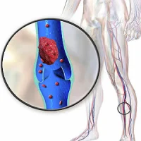
Cardiovascular disease is a leading cause of death worldwide. Bypass surgery or stent placement is often recommended in people with certain degrees of coronary arterial narrowing, or stenosis, but recent studies have shown that many of these patients do just as well with medical therapy. A key factor in treatment decisions is the haemodynamic significance of the lesion, meaning the degree to which the lesion is blocking blood from getting to areas of the heart.
A combination of invasive coronary angiography (ICA) and stress tests with single photon emission tomography (SPECT) myocardial imaging has been the gold standard for making these determinations, with ICA showing the blockages and SPECT the perfusion, or penetration of the blood into the tissue. However, ICA requires the use of a catheter that is threaded from a puncture point in the groin all the way up to the heart.
"Invasive angiography is generally safe, but it can cause vascular problems in a significant number of patients, most commonly at site of the puncture," says study author João A.C. Lima, MD, from Johns Hopkins Hospital and School of Medicine in Baltimore. "In rare cases, it can cause strokes or heart attacks. These risks are not trivial." The ICA/SPECT approach can also be expensive, as it often necessitates hospitalisation for the patient.
The current study aimed to determine if combined CT angiography (CTA) and CT myocardial stress perfusion imaging (CTP) could demonstrate similar or superior ability to ICA/SPECT in predicting future adverse events. Investigators compared the invasive and noninvasive approaches in 379 patients who were referred for ICA from November 2009 to July 2011. They looked at the ability of both techniques to predict whether or not a future major adverse cardiac event (MACE), such as a heart attack, revascularisation, arrhythmia or hospitalisation for chest pain or congestive heart failure would occur.
Overall, 51 patients experienced one or more major adverse cardiac events, including 49 revascularisations, five myocardial infarctions, one cardiac death, nine hospitalisations for chest pain or congestive heart failure, and one arrhythmia.
Both techniques had similarly high values for predicting MACE at two years after presentation and event-free survival, according to researchers. The two-year MACE-free rates for combined CT angiography and CT perfusion findings were 94 percent negative for coronary artery disease (CAD) versus 82 percent positive for CAD and were similar to combined ICA/SPECT findings (93 percent negative for CAD vs. 77 percent positive for CAD).
As both techniques are equally effective in identifying which patients "are going to have trouble down the road," Dr. Lima says, the noninvasive approach should be a preferred option for managing these patients because it is safer and less expensive.
Source: Radiological Society of North America
Image Credit: Pixabay
Latest Articles
CT angiography, heart attacks, stress tests, noninvasive imaging
A new study in the journal Radiology demonstrates the effectiveness of using noninvasive CT angiography and stress tests to detect which patients are likely to suffer a heart attack or other adverse cardiovascular event.