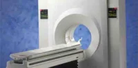
New nanoparticles developed by MIT chemists can simultaneously perform magnetic resonance imaging (MRI) and fluorescent imaging in living animals. These particles could assist scientists in monitoring a tumour's environment and determining whether drugs have successfully reached their targets.
An article appearing in Nature Communications describes how the particles, which carry distinct sensors for MRI and fluorescence, are able to track vitamin C in mice. Wherever there is a high concentration of vitamin C, the particles show a strong fluorescent signal but little MRI contrast. If there is not much vitamin C, MRI signal is strong but fluorescence is very weak.
The MIT researchers designed the particles so they can be assembled from building blocks made of polymer chains carrying either a fluorescent molecule called Cy5.5 or an organic MRI contrast agent called a nitroxide. When these building blocks are mixed together in a desired ratio, they form a specific nanosized structure the researchers call a branched bottlebrush polymer.
For this study, the particles were created with 99 percent of the chains carrying nitroxides and one percent carrying Cy5.5. Nitroxides are reactive molecules that contain a nitrogen atom bound to an oxygen atom with an unpaired electron. Nitroxides suppress Cy5.5's fluorescence, but when the nitroxides encounter a molecule such as vitamin C from which they can grab electrons, they become inactive and Cy5.5 fluoresces, the MIT team explained.
Future versions of the particles could be designed to detect reactive oxygen species that often correlate with disease, said Jeremiah Johnson, an assistant professor of chemistry at MIT and senior author of the study. They could also be customised to detect more than one molecule at a time. "You may be able to learn more about how diseases progress if you have imaging probes that can sense specific biomolecules," he added.
Extending Nitroxide Lifetime to Obtain Useful MRI images
Nitroxides typically have a very short half-life in living systems, but University of Nebraska chemistry professor Andrzej Rajca, a co-author of the study, recently discovered that their half-life can be extended by attaching two bulky structures to them. Incorporation of Prof. Rajca's nitroxide in Prof. Johnson's branched bottlebrush polymer architectures leads to even more improvements in the nitroxide lifetime. With these modifications, the research team noted, nitroxides can circulate for several hours in a mouse's bloodstream – long enough to obtain useful MRI images.
Prof. Johnson et al. reported these key findings:
The research team is now working to enhance the signal differences that they get when the sensor encounters a target molecule such as vitamin C. In addition, they have designed nanoparticles carrying the fluorescent agent plus up to three different drugs. This enables them to track whether the nanoparticles are delivered to their targeted locations. As Prof. Johnson pointed out: "That's the advantage of our platform – we can mix and match and add almost anything we want."
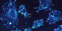
The imaging particles could also be used to assess the level of oxygen radicals in a patient's tumour, which can provide valuable information about how aggressive the tumour is.
For Steven Bottle, a professor of nanotechnology and molecular science at Queensland University of Technology, the most impressive element of the study is the combination of two powerful imaging techniques into one nanomaterial.
"I believe this should deliver a very powerful, metabolically linked, multi-combination imaging modality which should provide a highly useful diagnostic tool with real potential to follow disease progression in vivo," said Prof. Bottle, who was not involved in the study.
Source: ScienceDaily.com
Image Credit: Christine Daniloff/MIT
An article appearing in Nature Communications describes how the particles, which carry distinct sensors for MRI and fluorescence, are able to track vitamin C in mice. Wherever there is a high concentration of vitamin C, the particles show a strong fluorescent signal but little MRI contrast. If there is not much vitamin C, MRI signal is strong but fluorescence is very weak.
The MIT researchers designed the particles so they can be assembled from building blocks made of polymer chains carrying either a fluorescent molecule called Cy5.5 or an organic MRI contrast agent called a nitroxide. When these building blocks are mixed together in a desired ratio, they form a specific nanosized structure the researchers call a branched bottlebrush polymer.
For this study, the particles were created with 99 percent of the chains carrying nitroxides and one percent carrying Cy5.5. Nitroxides are reactive molecules that contain a nitrogen atom bound to an oxygen atom with an unpaired electron. Nitroxides suppress Cy5.5's fluorescence, but when the nitroxides encounter a molecule such as vitamin C from which they can grab electrons, they become inactive and Cy5.5 fluoresces, the MIT team explained.
Future versions of the particles could be designed to detect reactive oxygen species that often correlate with disease, said Jeremiah Johnson, an assistant professor of chemistry at MIT and senior author of the study. They could also be customised to detect more than one molecule at a time. "You may be able to learn more about how diseases progress if you have imaging probes that can sense specific biomolecules," he added.
Extending Nitroxide Lifetime to Obtain Useful MRI images
Nitroxides typically have a very short half-life in living systems, but University of Nebraska chemistry professor Andrzej Rajca, a co-author of the study, recently discovered that their half-life can be extended by attaching two bulky structures to them. Incorporation of Prof. Rajca's nitroxide in Prof. Johnson's branched bottlebrush polymer architectures leads to even more improvements in the nitroxide lifetime. With these modifications, the research team noted, nitroxides can circulate for several hours in a mouse's bloodstream – long enough to obtain useful MRI images.
Prof. Johnson et al. reported these key findings:
- Their imaging particles accumulated in the liver, as nanoparticles usually do.
- The mouse liver produces vitamin C, thus once the particles reached the liver, they grabbed electrons from vitamin C, turning off the MRI signal and boosting fluorescence.
- No MRI signal but a small amount of fluorescence was found in the brain, which is a destination for much of the vitamin C produced in the liver. In contrast, in the blood and kidneys, where the concentration of vitamin C is low, the MRI contrast was maximal.
The research team is now working to enhance the signal differences that they get when the sensor encounters a target molecule such as vitamin C. In addition, they have designed nanoparticles carrying the fluorescent agent plus up to three different drugs. This enables them to track whether the nanoparticles are delivered to their targeted locations. As Prof. Johnson pointed out: "That's the advantage of our platform – we can mix and match and add almost anything we want."
The imaging particles could also be used to assess the level of oxygen radicals in a patient's tumour, which can provide valuable information about how aggressive the tumour is.
For Steven Bottle, a professor of nanotechnology and molecular science at Queensland University of Technology, the most impressive element of the study is the combination of two powerful imaging techniques into one nanomaterial.
"I believe this should deliver a very powerful, metabolically linked, multi-combination imaging modality which should provide a highly useful diagnostic tool with real potential to follow disease progression in vivo," said Prof. Bottle, who was not involved in the study.
Source: ScienceDaily.com
Image Credit: Christine Daniloff/MIT
Latest Articles
MRI, nanoparticles, polymers, MIT, fluorescent imaging, nitroxide
New nanoparticles developed by MIT chemists can simultaneously perform magnetic resonance imaging (MRI) and fluorescent imaging in living animals. These pa...