At the International Society for Magnetic Resonance in Medicine’s (ISMRM) 21st Annual Meeting & Exhibition in Salt Lake City, Philips is showing a range of clinical functionalities in Magnetic Resonance Imaging (MRI).
An increasing global incidence of neuro and vascular disease across the world is a result of both ageing populations and increasingly unhealthy lifestyles. Philips is working in close collaboration with clinicians and researchers from across the world to develop new clinical functionalities that support high quality, clinically relevant diagnostics, whilst also improving patient throughput.
“Increasingly, MRI is proving its worth as a quick and cost effective diagnostic tool for the management of a wide variety of conditions; from stroke and dementia to tumour disease and trauma,” commented Dr. Jeroen Hendrikse, University Medical Center Utrecht, Department of Radiology. “A great example of this is pCASL, a functionality we’ve developed in close collaboration with the team at Philips. Not only does pCASL provide the user with an accurate assessment of brain perfusion - without the need for external contrast agent, a contraindication for many patients with renal insufficiency - but it’s also quick, taking just a short amount of additional time to scan.”
Dr Jeffrey Miller, MD, Pediatric Neuroradiologist, Phoenix Children’s Hospital, Arizona, adds, “Having access to these functionalities (pCASL, SWIp, mDIXON TSE and mDIXON Quant) means that we’re able to detect injuries that may not be apparent using conventional techniques, which can ultimately mean a better outcome for our patients. In particular, we’re seeing a real benefit from the image quality provided by the time-efficient mDIXON TSE technique in challenging anatomies such as spine, as well as the lesion delineation it provides.”
The functionalities*, developed collaboratively, that are being showcased at ISMRM include:
- pCASL (pseudo-Continuous Arterial Spin Labeling) – using arterial water as an endogenous tracer, pCASL provides cost effective and accurate assessment of changes in brain perfusion, without the need for contrast agent injection.
- SWIp** (Susceptibility Weighted Imaging with Phase difference) – utilizing phase information to enhance susceptibility contrast between tissues and a multi-echo acquisition to provide high SNR (signal-to-noise ratio), SWIp can help in the diagnosis of various brain lesions such as hemorrhage or venous malformation.
- mDIXON TSE – a unique and fast two-point DIXON technique that robustly separates water and fat signals, allowing for time-efficient, large FOV (field-of-view) fat-free imaging even in challenging areas such as head, neck or spine, and providing 4 contrasts in 1 scan.
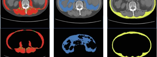
- mDIXON Quant** – utilizing an mDIXON multi-gradient-echo technique and state-of-the-art reconstruction for fast, volumetric, fat fraction quantification mapping, targeting anatomies such as the liver.
“As we continue to expand our MRI portfolio at Philips, our focus is to provide solutions that address the day-to-day needs of the radiology and clinical teams. Ultimately, innovation is only meaningful if it can bring about positive change for patients. Our commitment to achieving this is evident through our many and ongoing research collaborations,” commented Eric Jean, General Manager Global MR, Philips Healthcare. “It really is exciting to see that this approach is instrumental in addressing some of the most significant challenges in MRI today.”
* New functionalities available now on Philips Ingenia system, and on all Philips MRI systems by end of 2013
** 510(k) pending. Not available for sale in USA
Latest Articles
MRI, Philips
At the International Society for Magnetic Resonance in Medicine’s (ISMRM) 21st Annual Meeting & Exhibition in Salt Lake City, Philips is showing a r...


