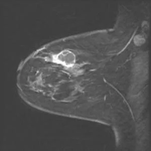
Microwave imaging can be used to monitor how well treatment for breast cancer is working, finds new research published in BioMed Central’s open access journal Breast Cancer Research. Microwave tomography, a low cost and repeatable technique, was able to distinguish between breast cancer, benign growths, and normal tissue.
Eight women with breast cancer were treated with chemotherapy until surgery. During treatment, magnetic resonance image was supplemented with microwave tomography at the Dartmouth-Hitchcock Medical Center in the U.S.. Regions of high conductivity corresponded to the tumours, low conductivity to normal tissues, and unlike other imaging techniques, body mass index (indicating the amount of body fat), age or breast density did not appear to affect the results.
Paul Meaney, Professor of Engineering at Dartmouth, who led the study explained, “By recalling patients for scans during their treatment we found that we could actually see tumours shrinking in women who responded to chemotherapy. Microwave tomography could therefore be used to identify women who are not responding to initial therapy and their treatment changed appropriately at an early stage.”
Image credit: Paul M Meaney
Latest Articles
Cancer, Imaging, Breast, Microwave
Microwave imaging can be used to monitor how well treatment for breast cancer is working, finds new research published in BioMed Central’s open access jo...


