New research suggests that a cheaper, better and safer way to look for signs of breast cancer may be with microwaves.
Neil Epstein, a NSERC CREATE I3T postdoctoral fellow at the University of Calgary in Canada and his colleagues -- engineering professor Paul Meaney of Dartmouth College's Thayer School of Engineering and Keith Paulsen, director of the Dartmouth Advanced Imaging Center and the Robert A. Pritzker Professor of Biomedical Engineering and Professor of Radiology at the Geisel School of Medicine at Dartmouth College, describe the microwave imaging system in the journal Review of Scientific Instruments.
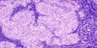
With the microwave imaging technique, the breast is suspended in a liquid bath and surrounded by an array of 16 antennae. Each antenna illuminates the breast individually with a low power microwave signal, while the other 15 antennae receive the signals transmitted through the breast. The process is repeated for all 16 antennae and provides data that can be used to produce a 3D representation of the breast, including the location of normal and cancerous tissue.
"The iterative image reconstruction algorithm computes what the dielectric property distribution must have been to generate the measured signal patterns," Epstein said. "It is quite similar to x-ray computed tomography, where the target is radiated from all of the surrounding directions and the data is synthesised to create an image of the internal structures."
The diagnostic screening systems that are used currently may be effective at detecting early signs of tumours, but they subject patients to radiation and discomfort because of breast compression. The microwave imaging technique does not compress the breast and while it may not provide the spatial resolution, it does offer better specificity.
Once tumours are localised, this new technique can prove to be more adept at identifying if a tumour is benign or malignant. Thus the microwave imaging system can potentially solve the specificity problem that is associated with conventional imaging techniques. Epstein believes that microwave imaging could fill this niche in combination with other modalities.
Source: American Institute of Physics
Image Credit: N.Epstein/U.Calgary