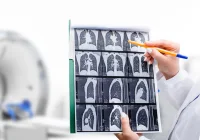
Magnetic resonance imaging (MRI) plays a vital role in the detection and characterisation of liver lesions, particularly through T1-weighted imaging during various contrast-enhanced phases. While these techniques offer rich diagnostic data without ionising radiation, they face practical limitations, notably the requirement for prolonged breath-holds. These limitations can impair image quality due to motion artefacts and reduce diagnostic confidence.
Recent advances in artificial intelligence have led to the development of reconstruction techniques that may improve image quality and shorten scan duration. A study published in European Radiology Experimental evaluated the efficacy of an AI-augmented T1-weighted hepatobiliary-phase liver MRI sequence in comparison to standard imaging methods, with a focus on image quality and lesion detectability.
Improving Image Quality with AI-Augmented Reconstruction
The study involved fifty patients undergoing gadoxetate-enhanced liver MRI, who were imaged using both standard and AI-augmented T1-weighted sequences. Two sequences were used: a routine 17-second breath-hold and an accelerated 12-second breath-hold with reduced phase resolution. Each was reconstructed using standard methods, neural network (NN)-only, iterative denoising (ID)-only and a combined NN+ID approach.
Quantitative analysis revealed that the 17-second NN+ID images had a significantly higher liver-to-portal vein contrast-to-noise ratio (CNR) compared to both the standard and NN-only reconstructions. Qualitatively, both the 17- and 12-second NN+ID images scored higher than the standard reconstruction for overall image quality, CNR and edge sharpness of vessels and lesions. These results underscore the potential of AI-augmented reconstruction to enhance visual clarity, reduce artefacts and improve diagnostic confidence without extending scan time.
Must Read: Improving Upper Abdominal MRI with DL Reconstruction
Maintaining Diagnostic Accuracy with Shorter Breath-Holds
Shorter breath-hold durations are particularly beneficial for patients with limited respiratory capacity. The study showed that reducing the acquisition time from 17 to 12 seconds using the NN+ID approach did not compromise image quality. Both versions performed similarly across all measured parameters, including sharpness, CNR and susceptibility to respiratory motion artefacts. While one of the two readers observed fewer motion artefacts in the NN+ID series, the discrepancy in assessment between the two readers was attributed to individual interpretation and experience.
Importantly, lesion detection rates were comparable between the 12-second NN+ID and the 17-second standard reconstructions. There was no significant difference in the number of lesions identified or the size of the smallest detectable lesion. These findings suggest that AI augmentation not only preserves diagnostic integrity but also allows more efficient and patient-friendly imaging, potentially leading to wider clinical adoption.
Study Strengths, Limitations and Clinical Implications
This study demonstrated the feasibility and advantages of incorporating AI-based reconstruction into liver MRI protocols. By significantly improving image quality without requiring longer breath-holds, this technique holds particular promise for populations such as elderly patients or those with respiratory limitations. Furthermore, consistent performance across quantitative and qualitative metrics indicates that AI-augmented sequences could standardise and enhance imaging outputs.
However, limitations must be acknowledged. The study population consisted solely of patients with colorectal cancer and was confined to a single imaging centre using one MRI system type. Additionally, only hepatobiliary-phase images were assessed, meaning potential benefits in other imaging phases remain unverified. The study also did not evaluate the specificity of lesion characterisation nor the clinical relevance of lesions detected, as benign and malignant findings were analysed together without further stratification.
Despite these constraints, the outcomes reinforce prior research supporting the clinical utility of deep learning in MR imaging. AI-driven image reconstruction not only addresses key technical challenges such as acquisition time and image artefacts but also aligns with the growing demand for efficient, high-quality imaging in oncology and beyond.
AI-augmented reconstruction, combining neural network interpolation with iterative denoising, significantly enhances image quality in hepatobiliary-phase liver MRI while enabling reduced breath-hold durations. This approach maintains diagnostic accuracy in lesion detection and has the potential to improve patient comfort and compliance. While further studies are needed to validate its effectiveness across diverse patient groups and imaging systems, the findings support broader clinical integration of AI tools into routine liver MRI protocols.
Source: European Radiology Experimental
Image Credit: iStock


