Scientists in the UK have used an imaging technique, that can accurately measure the distance between two protein molecules, as a new way to identify patients who could benefit from certain breast cancer treatments.
In a study published in the journal Oncotarget, the researchers from King’s College London, in collaboration with scientists at Cancer Research UK/MRC Oxford Institute for Radiation Oncology, used fluorescence lifetime imaging to measure the distance between HER2 and HER3 proteins in breast cancer cells from patients.
The researchers think that patients whose imaging results show that these proteins have joined together could benefit from HER2 targeted treatment, regardless of whether their tumour has high levels of HER2.
HER2 is a protein which can cause cancer cells to grow. HER2-positive breast cancer cells have high levels of the protein and can be targeted with drugs that block its effects and stop the cancer from growing -- drugs being used now include Herceptin and Tykerb. Patients who could benefit from these drugs are identified by testing their cancer cells to see if they show high levels of the HER2 protein.
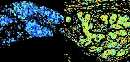
(ABOVE) The left image is a tissue sample with low levels of HER2 and HER3 bonded together. The right is a sample with high levels.
But this imaging technique, carried out in tumour cells, could pick up additional patients in the future who would respond well to HER2-targeting drugs. It could also confirm which patients may not be suitable for these treatments.
This test could help predict which drugs won’t work in patients and avoid prescribing unnecessary treatments, not only to improve treatment for breast cancer but also for other cancers -- including bowel and lung cancer, explained lead author, Professor Tony Ng, at King’s College London and University College London.
Story Source: Cancer Research UK.
Journal Reference:
1. Weitsman et al. HER2-HER3 dimer quantification by FLIM-FRET predicts breast cancer metastatic relapse independently of HER2 IHC status. Oncotarget, July 2016
See also:Combining Imaging And Maths in BRIM Breast Cancer Biomarker