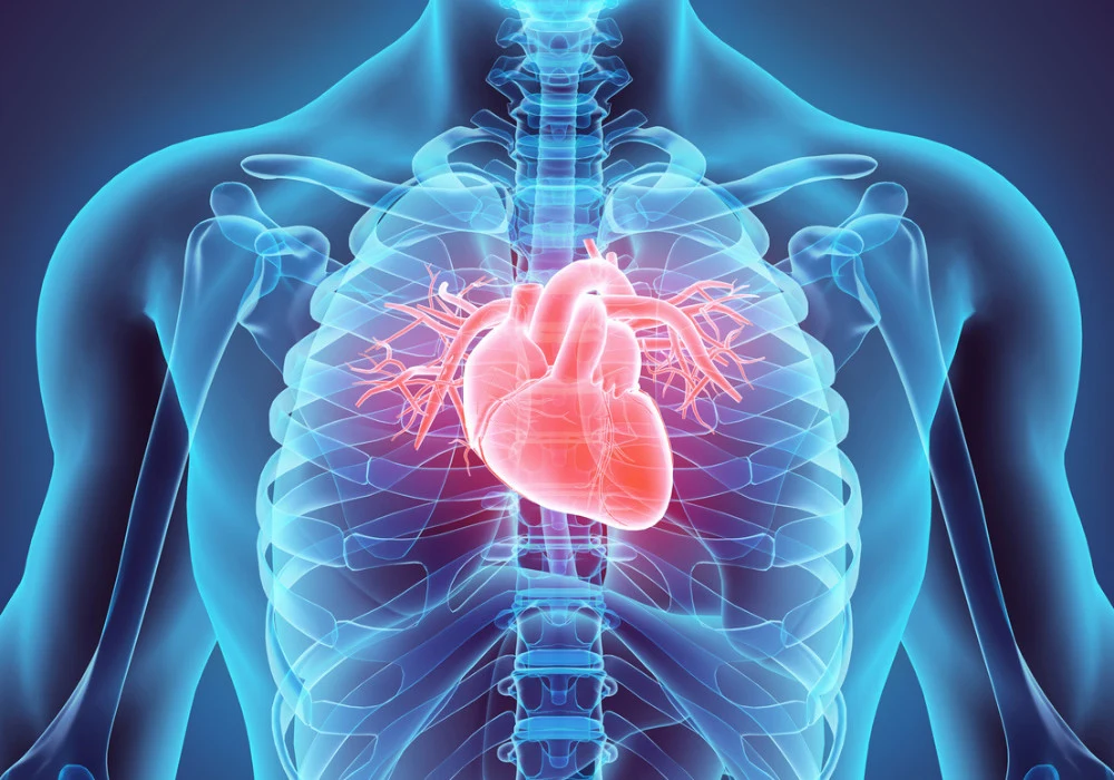
Cardiac imaging modalities such as computed tomography (CT) and magnetic resonance imaging (MRI) have transformed the diagnosis and management of heart diseases. These imaging techniques offer high-resolution images, helping clinicians detect cardiovascular abnormalities non-invasively. While the primary goal of these modalities is to assess heart conditions, they often capture parts of surrounding structures, leading to incidental findings beyond the heart, known as extracardiac findings. Clinically relevant extracardiac findings, defined as findings that necessitate follow-up or influence management, are particularly significant. A recent review published by Radiology: Cardiothoracic Imaging explores the prevalence and implications of such findings based on data from the European MR/CT registry, which includes over 400,000 cardiac CT and MRI examinations.
Prevalence of Clinically Relevant Extracardiac Findings
The European Society of Cardiovascular Radiology's MR/CT registry reveals that clinically relevant extracardiac findings are common in cardiac imaging. The overall prevalence at cardiac CT examinations is 3.28%, while at MRI, it is 1.50%. These numbers highlight the potential for detecting incidental pathologies in areas such as the lungs, abdomen, and bones, among others. In CT, most extracardiac findings occur in the lungs, including pulmonary lesions and effusions, while abdominal lesions and enlarged lymph nodes are also frequently observed. MRI findings are similar but less frequent, often involving pleural effusion, pulmonary nodules, and abdominal abnormalities.
Interestingly, the prevalence of extracardiac findings varies depending on the imaging modality and the patient's examination indication. Cardiac CT scans performed for transcatheter aortic valve replacement (TAVR) procedures show the highest rate of extracardiac findings, owing to the broader area of coverage that extends into the abdomen and pelvis. This expanded imaging field increases the chance of detecting incidental findings, particularly in older populations. In contrast, MRI scans for myocarditis and structural heart disease reveal fewer extracardiac findings but still demonstrate clinically significant detection rates, underlining the broader diagnostic value of cardiovascular imaging.
Factors Influencing the Detection of Extracardiac Findings
Several factors influence the likelihood of detecting extracardiac findings during cardiac imaging, with patient age being a significant predictor. The registry data show that for both CT and MRI, the prevalence of extracardiac findings increases with age, with a 2% increase for each additional year. This association is particularly evident in older populations undergoing TAVR, where the median age is significantly higher, resulting in more frequent detection of lung and abdominal lesions. Additionally, structural heart disease and older age groups are linked with a higher prevalence of these findings. This is not surprising, as age-related changes in the body often manifest as extracardiac pathologies such as pulmonary nodules or abdominal abnormalities.
The type of examination indication also plays a crucial role. For instance, CT scans conducted for coronary artery disease (CAD) typically focus on the heart and surrounding vessels, limiting the detection of extracardiac findings. In contrast, when the imaging scope is expanded to evaluate structural heart diseases or TAVR planning, the prevalence of incidental findings increases due to the larger field of view. MRI examinations, which often focus on soft tissue imaging, tend to reveal extracardiac findings related to the lungs and lymph nodes, further broadening the diagnostic capabilities of these modalities.
Clinical Implications of Extracardiac Findings
The discovery of extracardiac findings during cardiac imaging has significant clinical implications. Some of these findings, such as pulmonary embolism or malignancies, require urgent follow-up and treatment. Others, like benign lesions, may not immediately impact the patient's health but still warrant further observation. The ability to identify extracardiac pathologies early on can lead to timely interventions, potentially improving patient outcomes and preventing future complications. However, the challenge lies in distinguishing between clinically relevant and irrelevant findings. Misinterpretation of benign lesions as malignant can lead to unnecessary follow-up tests, increasing healthcare costs and causing patient anxiety.
Radiologists are uniquely positioned to assess these extracardiac findings given their expertise in interpreting cross-sectional imaging. Their role is not limited to evaluating the heart but extends to adjacent structures that may be included in the imaging field. This requires a comprehensive understanding of cardiovascular and non-cardiovascular pathologies, which further emphasises the importance of multidisciplinary collaboration in patient care. The European MR/CT registry data supports that detecting and interpreting extracardiac findings is crucial for holistic patient management, ensuring that incidental findings are appropriately addressed to improve long-term health outcomes.
Extracardiac findings are a common and clinically relevant byproduct of cardiac imaging, with CT and MRI revealing significant incidental pathologies beyond the heart. The European MR/CT registry highlights the significance of these findings, particularly in older patients and those undergoing broader imaging examinations like TAVR. As the use of cardiac imaging continues to rise, so will the detection of extracardiac pathologies, underscoring the need for careful interpretation by radiologists. Identifying and managing these incidental findings can lead to earlier interventions, improving patient care and preventing potential complications. Ultimately, cardiac imaging serves to diagnose heart conditions and contributes to a more comprehensive understanding of the patient's overall health.
Source: Radiology
Image Credit: iStock


