


The IRCCS in Bologna has inaugurated a state-of-the-art integrated PET/CT system. This cutting-edge technology allows for the entire human body to be studied in a single scan, even detecting the smallest tumour cells. In addition to reducing examination duration from 12 minutes to less than a minute, the new device offers unique opportunities for research and development in oncology, particularly in the field of radiopharmaceuticals. The Nuclear Medicine department at Sant'Orsola is already renowned for the quality of its scientific studies, and this latest investment will further enhance its reputation. The investment forms part of the wider research and development plan for the Policlinic, which includes the purchase of large diagnostic equipment.
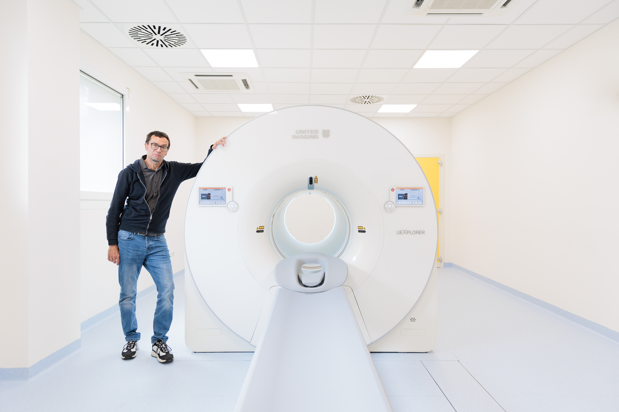
BOLOGNA. For the first time in Europe, researchers can track the distribution of a molecule throughout the human body with a single scan. This is made possible by the new uEXPLORER, the World's First Total-Body PET/CT scanner', which was inaugurated May 20th at the Nuclear Medicine Unit of the IRCCS Azienda Ospedaliero Universitaria di Bologna - Policlinico di Sant'Orsola. The investment has enabled the development of a technology that drastically shortens examination times and increases the ability to detect tumour micro-metastases. This opens up important research prospects for the development of new drugs for both the diagnosis and treatment of tumours.
The device completes the technological endowment in the field of advanced imaging dedicated to research (RM3 tesla and Spectral CT), enabling the Bologna IRCCS to make a further qualitative leap forward and confirm itself as a reference point at the international level. The 'Total Body PET/CT' technology, co-developed by UC Davis and United Imaging Healthcare, and distributed exclusively nationwide by FORA, is arriving for the first time on the European scene. Currently, there are only approximately 20 systems of this type installed worldwide.
Innovations: Almost Two-Metre Field of View and Ten Times Greater Sensitivity
The uExplorer 'Total-Body PET/CT' overcomes the limitations of conventional devices, which are characterised by a limited field of view and sensitivity. The PET/CT scanners currently in use can only scan circumscribed segments of the human body, typically between 15 and 30 centimetres. This means that, until now, several scans had to be taken by moving the couch inside the machine to obtain a full-body image. The 'Total-Body PET/CT', in contrast, boasts an axial field of view of 194 centimetres and can therefore examine the entire human body in a single scan. In addition, the new device is characterised by a detection sensitivity that is ten times higher than standard.
From Photo to Video: The Technology to Track Radiopharmaceuticals in Real-Time
Thanks to the almost two-metre field of view and higher sensitivity, as well as highly advanced reconstruction algorithms and post-processing software including Deep Learning and Artificial Intelligence techniques, the new device also introduces the possibility of dynamic full-body studies with frames of the order of a second.
From the static images of conventional devices, we then move on to the possibility of following the path and distribution of radiopharmaceuticals in dynamic mode, observing their evolution over time. In practice, this represents a shift from photography to cinema.
The New Frontiers of Research
This feature represents a significant advancement for research. The new Total-Body PET/CT offers a unique opportunity to study the pharmacokinetics and biodistribution of whole-body tracers, opening up important prospects for the development of new radiopharmaceuticals for both diagnostic and therapeutic purposes. The study activity will focus on the oncological field, with a particular focus on pathologies that cannot be examined with FDG tracer, such as kidney cancer. It will also examine inflammatory processes and autoimmune diseases.
The device's unique capabilities are particularly well-suited to competitive research calls, such as European ones, and to collaborations with various partners for the development of innovative approaches.
Benefits and Clinical Applications: An Extra Tool Against Myelomas and Lymphomas (and More)
The main clinical and research applications include the identification of tumour residues (e.g. in myelomas and lymphomas), the diagnosis of neoplastic recurrences (breast tumours, melanomas, CNS tumours, digestive tract tumours and ovarian tumours), the study of infectious processes (endocarditis and septicaemia) and the early diagnosis of treatment response (CAR-T, immunotherapy, target therapy).
Thanks to its high sensitivity, the device also enables a reduction in the administered radiopharmaceutical dose by more than 80%, thus limiting the radiation dose to which patients and operators are subjected on average. The dose is just 0.002 milliCuries per kilogram (compared to the standard average of 0.8/0.15 mCi/kg). This is a significant advantage, particularly for paediatric and young patients who have to repeat the examination several times.
Finally, the ability to scan the entire human body in a single scan allows for a reduction in examination duration from 12 minutes to less than one minute. This represents a significant time saving, which translates into greater comfort for patients, particularly those suffering from severe pain (e.g. in the presence of a neoplastic bone disease). It also reduces the need for sedation in paediatric patients or patients with limited ability to cooperate.
Investments to Strengthen Oncology Research at the Polyclinic
The arrival of the new PET/CT is part of a much broader framework of development for research purposes of the company's diagnostic devices. Since 2020, large pieces of equipment have been installed in the halls of the IRCCS Policlinico di Sant'Orsola at a total cost of almost EUR 17 million. The sum includes the long-term rental or purchase of two computerised tomography, a surgical robot, a linear accelerator, a spectral CT (an integrated PET/CT system), a high-field magnetic resonance tomograph and two digital mammographs with tomosynthesis.
The Policlinico di Sant'Orsola is also in the process of renewing its PNRR-related technologies for which it is on schedule.
Nuclear Medicine at Sant'Orsola is Among the Top Five in Europe
The new Total-Body PET/CT is in addition to the four PET/CT tomographs and the radiopharmacy with a 17 MeV cyclotron for the in-house production of both standard and innovative radiopharmaceuticals. Thanks to this complex instrumentation, Bologna Nuclear Medicine is one of the very few centres capable of working on both clinical and research activities on different isotopes.
For many years, the centre has been the most significant scientific player in Italy in this field, publishing more than 60 articles per year in major scientific journals. Professor Stefano Fanti, the director of the centre, is a leading expert in the field and has an H-Index of 98 on Google Scholar.
The Nuclear Medicine department of the IRCCS Azienda Ospedaliero-Universitaria di Bologna, which provides services at both the Sant'Orsola General Hospital and the Maggiore Hospital, is a leading international reference point for both clinical activity and research. PET is the centre's flagship service, with over 14,000 procedures performed annually. The IRCCS is the largest PET centre in Italy and one of the top five PET centres in Europe. The activity has been consistently growing over the years, with no significant slowdown even during the Covid period. To this day, it is still able to guarantee compliance with waiting times.
Oncological Research at Sant'Orsola with Great Equipment
In 2020, the Policlinico di Sant'Orsola was granted IRCCS (Istituto di Ricovero e Cura a Carattere Scientifico) status in the fields of oncology and organ transplantation/insufficiency. Among the platforms of the IRCCS, the one dedicated to advanced imaging is already operational, offering a 3-tesla magnetic resonance imaging and a spectral computed tomography. The PET/CT Total-Body completes the technological endowment of advanced imaging tools.
What is PET/CT?
Positron Emission Tomography (PET) is the most effective molecular imaging method currently available, offering unparalleled specificity, sensitivity and accuracy. This diagnostic technique employs the use of a radiopharmaceutical and a tomograph, which are capable of detecting and tracking the radiopharmaceutical within the body.
The radiopharmaceutical is administered intravenously and consists of a radioactive isotope stably bound to a carrier molecule, such as glucose, which carries it within the body. The tracer is distributed selectively in the areas to be studied, making them visible through the tomograph's detection of radioactivity. This enables the examination to provide valuable insights into the distribution and characteristics of the pathology.
PET scans can detect changes in tissue metabolism, providing functional information about the areas of the body being analysed. This technology offers many advantages over other imaging methods, including the ability to monitor tumour lesions, assess treatment response and identify the potential for metastasis at an early stage.
Integrated PET/CT systems combine PET features with anatomical information that can be detected using the latest generation of computerised tomography (CT). This hybridisation allows for the acquisition of a complete clinical picture, as well as precise information on the nature, stage and evolution of the disease during and after treatments.
Source & Image Credit: United Imaging Healthcare