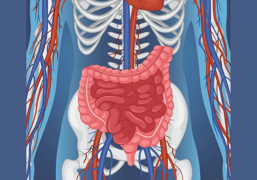
Surgical intervention in Crohn’s disease (CD) often stems from complications caused by intestinal fibrosis, a condition marked by excessive connective tissue formation in the bowel wall. Accurate assessment of this fibrosis remains a clinical challenge, as current diagnostic methods either lack precision or involve invasive procedures. With no universally accepted gold standard for fibrosis evaluation, researchers continue to seek non-invasive yet reliable alternatives. The integration of unenhanced spectral CT with 3D-printing technology presents a novel approach for visualising and quantifying fibrotic changes. By enabling precise layer-to-layer correspondence between imaging and pathological specimens, this method could significantly improve the preoperative diagnosis of fibrosis in CD patients.
Spectral CT Imaging and Parameter Correlation
Spectral CT enterography (CTE) provides multi-parametric imaging, generating various data points such as electron density (ED), Hounsfield units (HU), and energy spectrum curve slopes (λ1 and λ2). Among these, ED and HU at MonoE 50 keV were most significantly correlated with histologically confirmed fibrosis. Through statistical analyses, it was demonstrated that these parameters not only followed normal distribution but also maintained strong correlations even after accounting for the confounding influence of inflammation. Using LASSO regression, ED and HU at 50 keV were identified as key predictors, leading to the development of a diagnostic model capable of differentiating between non-mild and moderate-severe fibrosis. The model showed promising performance in the training set, with an area under the curve (AUC) of 0.828, sensitivity of 77.3%, and specificity of 82.6%.
Must Read: Imaging Techniques for Crohn Disease and Ulcerative Colitis
Further performance validation was conducted using an independent dataset, where the model maintained strong diagnostic accuracy (AUC of 0.812), particularly excelling in specificity (89.7%). After excluding inflammation as a variable, the model's AUC rose to 0.933, suggesting that fibrosis-specific markers remained robust even under inflammatory conditions. This demonstrates the model’s potential for consistent performance in real-world clinical settings where inflammation and fibrosis often co-occur.
Enhancement through 3D-Printing and Layer Matching
A major innovation in this study was the application of 3D printing to support exact anatomical alignment between imaging and pathological specimens. A custom-designed intestinal mold was fabricated using CT data, ensuring that resected bowel segments could be sectioned in precise correspondence with preoperative imaging. This allowed for highly accurate cross-validation of imaging markers with full-thickness histopathological slides.
By establishing a one-to-one match between radiological and pathological layers, the study revealed enhanced correlations between imaging parameters and fibrosis scores. When the same parameters were evaluated without 3D-printed correspondence, diagnostic performance dropped. Notably, parameters such as HU at multiple keV levels lost statistical significance or showed diminished correlation strength. This confirmed that spatial heterogeneity in CD lesions could be better understood and evaluated through exact anatomical matching, which would otherwise be missed in conventional imaging-pathology comparisons.
The added precision brought by 3D-printing not only elevated diagnostic performance but also increased confidence in parameter-based prediction models. This methodological refinement suggests a broader role for 3D-printing in imaging-guided diagnostics, particularly in diseases where tissue structure and distribution vary significantly, as is often the case in CD-related fibrosis.
Clinical Implications and Model Accessibility
The clinical utility of the diagnostic model is underscored by its accessibility. A web-based dynamic nomogram was created, enabling clinicians to input ED and HU values to instantly obtain fibrosis risk assessments for specific intestinal segments. This tool offers an easy-to-use and radiation-free option for ongoing disease monitoring and decision-making, which is particularly valuable in managing chronic conditions like CD.
The non-contrast nature of spectral CT in this study further supports patient safety, reducing the need for contrast-enhanced scans and associated risks. This is especially beneficial for CD patients, who often undergo repeated imaging throughout their lifetime due to the disease’s relapsing nature. The ability to distinguish fibrosis from inflammation without contrast enhancement aligns well with the growing emphasis on low-burden diagnostic techniques.
Moreover, the model's reliability under inflammatory conditions reinforces its clinical relevance. Inflammation and fibrosis often coexist, complicating treatment decisions. By demonstrating diagnostic stability even after adjusting for inflammatory scores, the model offers a tool that is robust in complex pathological scenarios. This reliability may assist in guiding surgical decisions, potentially reducing unnecessary resections and improving patient outcomes.
The integration of unenhanced spectral CT parameters with 3D-printing technology represents a significant advancement in the assessment of intestinal fibrosis in Crohn’s disease. This novel approach offers a reproducible, accurate, and minimally invasive method for evaluating the extent and severity of fibrosis, addressing longstanding diagnostic limitations. The strong correlation between imaging metrics and histological fibrosis, enhanced through anatomical co-registration, confirms the value of combining these technologies.
With high diagnostic performance, particularly when inflammation is controlled, the developed model has the potential to influence treatment strategies by identifying patients more likely to benefit from surgical intervention. The inclusion of a dynamic online nomogram further translates research findings into practical tools for clinical application. While future studies with larger and more diverse cohorts are necessary, the current findings underscore a promising path toward personalised and precision-based care for Crohn’s disease patients.
Source: Insights into Imaging
Image Credit: Freepik


