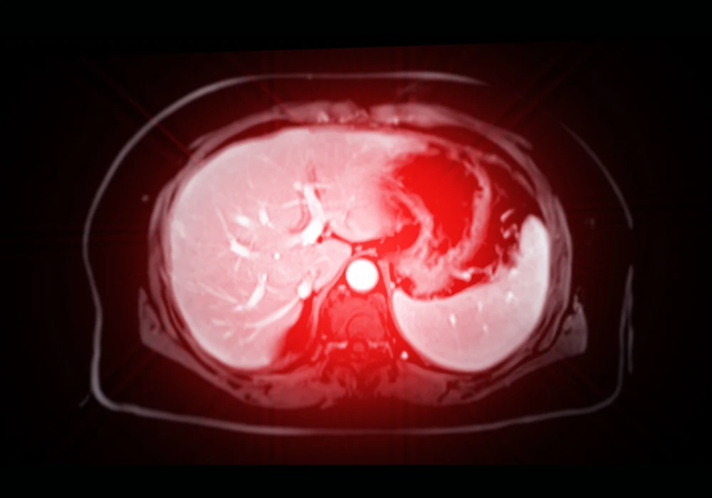
Hepatocellular carcinoma (HCC) is a major global health burden and ranks among the leading causes of cancer-related deaths. Despite curative treatments such as liver resection or transplantation, HCC exhibits high recurrence rates, often due to microvascular invasion (MVI), a pathological hallmark of tumour aggressiveness. Accurate preoperative prediction of MVI is crucial for optimising surgical planning and stratifying recurrence risk. Currently, MVI can only be diagnosed through pathological analysis after surgical resection.
Non-invasive imaging techniques like MRI have shown potential in predicting MVI, particularly when integrated with deep learning methods. However, many existing models suffer from limited generalisability, often due to reliance on single-centre data. To overcome this, a multicentre study developed an interpretable adversarial network-based deep learning (AD-DL) model using MRI data from five centres, aiming to deliver robust MVI prediction and prognostic stratification.
Model Development and Dataset Composition
The study retrospectively analysed MRI and clinical data from 546 patients diagnosed with HCC, drawn from five tertiary medical centres. Patient data from three centres were split into training and internal test sets, while two other centres provided external test cohorts. Additionally, transcriptomic data from The Cancer Imaging Archive (TCIA) were used for biological interpretability analysis. The MRI scans followed protocols aligned with AASLD guidelines, and tumour regions were manually segmented across seven MRI sequences: in-phase (IP), opposed-phase (OP), T2-weighted imaging (T2WI), non-contrast phase (NCP), arterial phase (AP), portal venous phase (PVP) and delayed phase (DP).
A clinical-radiological (CR) model was first built using features identified through univariate and multivariate logistic regression. The model included alpha-fetoprotein (AFP) levels, intratumoral vascularity and peritumoral arterial hyperenhancement. Although the CR model demonstrated reasonable internal performance, its accuracy and area under the curve (AUC) declined in external validation.
To enhance generalisability, the AD-DL model was designed using adversarial training strategies. The architecture included a feature extraction module, MVI classifier, hospital classifier and a gradient-reversal layer (GRL) to learn domain-invariant features. The model tested various MRI sequence combinations, with the IP, OP and AP sequence set showing the highest predictive performance.
Performance and Comparative Evaluation
The CR model achieved an internal test set AUC of 0.759 but performed less effectively in external tests, recording AUCs of 0.636 and 0.721. The SE-DL model, which did not use adversarial training, improved performance slightly but still fell short in generalisability. In contrast, the AD-DL model consistently outperformed both the CR and SE-DL models. In the internal test set, it reached an AUC of 0.793; in external test sets 1 and 2, it achieved AUCs of 0.801 and 0.773 respectively. Sensitivity, accuracy and specificity metrics also favoured the AD-DL model, especially in external settings.
Must Read: Consensus Imaging Guidelines for HCC Treatment
The AD-DL model's predictions were significantly associated with early recurrence-free survival (ERFS). Among patients in the Taizhou centre, those identified as MVI-positive by the model had a mean ERFS of 14.1 months compared to 20.7 months in the MVI-negative group. Grad-CAM visualisation showed that the AD-DL model focused on tumour margins for MVI-positive predictions, aligning with known pathological observations that most MVI occurs peritumorally.
Biological Function Analysis and Interpretability
To support model interpretability, the study conducted bioinformatics analysis using transcriptomic data from TCIA. A total of 198 differentially expressed genes (DEGs) were identified between MVI-positive and MVI-negative groups as predicted by the AD-DL model. Gene ontology analysis revealed enrichment in metabolic processes and synaptic regulation, while KEGG pathway analysis highlighted involvement of the Wnt and Hippo signalling pathways.
Protein–protein interaction (PPI) network analysis identified hub genes such as Bmp4, which has been linked to endothelial cell migration. Immune cell profiling using the CIBERSORT algorithm further showed increased infiltration of regulatory T cells and M0 macrophages in MVI-positive tumours, while monocytes and M2 macrophages were reduced. These findings suggest a link between model predictions and biologically relevant tumour microenvironment features.
The study demonstrated that an adversarial network-based deep learning model trained on MRI data can robustly predict microvascular invasion in hepatocellular carcinoma. Compared to clinical-radiological and non-adversarial deep learning models, the AD-DL model showed superior accuracy, generalisability and correlation with clinical outcomes. Grad-CAM and bioinformatics analyses provided insights into the biological underpinnings of model predictions, enhancing interpretability and clinical trust. The integration of domain generalisation techniques and transcriptomic validation marks a significant advancement in non-invasive MVI assessment. Prospective, multicentre studies are now needed to confirm these findings and support broader clinical adoption.
Source: Insights into Imaging
Image Credit: iStock


