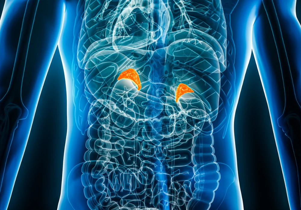
Adrenal adenomas represent the most frequent type of adrenal lesion, accounting for up to 80% of cases. Accurate identification is vital, as management pathways differ markedly between benign adenomas and other adrenal pathologies. Unenhanced computed tomography has long been the standard tool for detecting lipid-rich adenomas, with a 10 Hounsfield unit (HU) threshold commonly applied to distinguish adenomas from non-adenomas. While effective on conventional imaging, this threshold does not necessarily translate to newer technologies. Photon-counting CT (PCCT), a recent advance in imaging, enables the generation of virtual noncontrast (VNC) images from contrast-enhanced acquisitions. This technique reduces the need for separate noncontrast scans, lowering both radiation exposure and cost. However, its diagnostic performance in adrenal imaging and the thresholds required for reliable differentiation have remained uncertain. A prospective evaluation has now clarified the value and limitations of VNC reconstructions in this setting.
Diagnostic Value of VNC Imaging
The prospective analysis enrolled 54 participants with a mean age of 45 years, comprising 68 adrenal lesions in total. Fourteen participants had bilateral lesions, and the mean lesion size was 15 mm. Of the 68 lesions, 34 were identified as adenomas through pathology, attenuation values on true noncontrast imaging, or clinical stability with endocrine exclusion of malignancy. The remaining 34 lesions were non-adenomas, including pheochromocytoma and adrenal hyperplasia.
Must Read: Dual-Space Randomisation in Abdominal Segmentation
PCCT scans were performed using a dual-source system, with both unenhanced and portal venous phase acquisitions. VNC images were reconstructed with two algorithms: conventional VNC (VNCConv) and PureCalcium VNC (VNCPC). Attenuation was assessed using both two-dimensional single-slice measurements and semiautomatic three-dimensional segmentation. Across all lesions, VNC images consistently showed higher attenuation than true noncontrast (TNC) images. Mean differences reached 8.8 HU for VNCConv and 6.9 HU for VNCPC, confirming systematic overestimation. Despite this, values between 2D and 3D measurements did not differ significantly, suggesting that single-slice measurement at the largest diameter is sufficient in routine practice.
Radiomic analysis added further insights. Agreement between TNC and VNC was excellent for shape features, with more than 60% achieving an intraclass correlation coefficient (ICC) above 0.8. First-order and texture features showed only moderate consistency, with ICCs averaging 0.671 and 0.616 respectively. This indicates that while VNC can replicate the gross morphology of adrenal lesions, finer intensity and texture characteristics may not be reproduced with equal reliability.
Thresholds and Diagnostic Accuracy
When comparing adenomas and non-adenomas, marked differences emerged in attenuation values across imaging types. Adenomas measured a mean of 7.8 HU on TNC but were substantially overestimated on VNCConv (18.7 HU) and VNCPC (16.4 HU). Non-adenomas showed higher baseline values, averaging 28.2 HU on TNC, 33.3 HU on VNCConv, and 31.1 HU on VNCPC. These shifts underline the need to adjust thresholds when interpreting VNC images.
True noncontrast imaging achieved the highest diagnostic performance, with an area under the curve (AUC) of 0.914 and an optimal threshold of 17 HU, producing 88% sensitivity and 82% specificity. VNCConv reached an AUC of 0.841, with an optimal cut-off of 26 HU yielding balanced sensitivity and specificity of 79%. VNCPC showed a similar AUC of 0.838, with 18 HU as the optimal threshold, achieving 62% sensitivity and 91% specificity. Importantly, the established 10 HU threshold performed poorly in VNC, reducing sensitivity to as low as 18%.
Proposed practical thresholds were therefore set higher: 25 HU for VNCConv and 20 HU for VNCPC. At these levels, sensitivities and specificities approached those of TNC, though not surpassing it. Stratification by lesion size revealed further nuances. Smaller lesions benefited most from VNC-based thresholds. Lesions under 10 mm achieved sensitivities and specificities exceeding 90% when using VNCPC at 20 HU. By contrast, performance declined in lesions over 15 mm, where accuracy dropped to 56%. This indicates that VNC imaging is particularly reliable in small-volume disease, but less so in larger masses where heterogeneity may confound attenuation values.
Clinical Implications and Efficiency
The findings carry clear clinical implications. Many adrenal lesions are discovered incidentally on single-phase contrast-enhanced CT, where no true noncontrast data are available. In such cases, VNC imaging generated from PCCT offers a practical solution, avoiding repeat scans and reducing both radiation exposure and healthcare costs. Adjusted thresholds of 25 HU for VNCConv and 20 HU for VNCPC provide clinicians with guidance on interpreting these reconstructions more reliably than relying on the conventional 10 HU rule.
From a workflow perspective, the observation that 2D single-slice attenuation correlates well with 3D volumetric measurements has important efficiency benefits. Radiologists can obtain clinically relevant values without extensive segmentation, saving time and resources. Radiomic analysis also highlights the strengths and limitations of VNC. While shape features are preserved with high fidelity, variability in texture and first-order features suggests that VNC should be interpreted with caution when more complex radiomic modelling is required.
The study also found that lesion size itself was not significantly different between adenomas and non-adenomas, limiting its value as a discriminating parameter. Size thresholds performed poorly, with AUC values around 0.59. This underscores the central role of attenuation thresholds in adrenal imaging, whether measured on TNC or VNC images.
Photon-counting CT expands the possibilities of adrenal imaging by enabling reliable virtual noncontrast reconstruction. While VNC overestimates attenuation compared with true noncontrast images, adjusted thresholds improve diagnostic accuracy. With values of 25 HU for VNCConv and 20 HU for VNCPC, clinicians can more accurately distinguish adenomas from other adrenal lesions, particularly in smaller lesions where diagnostic accuracy is highest. True noncontrast imaging remains the gold standard, but in routine clinical practice, VNC reconstructions offer a valuable alternative, reducing the need for additional scans and minimising radiation exposure. Adapting to these new thresholds will be key to maximising the benefits of photon-counting CT in adrenal lesion management.
Source: European Radiology Experimental
Image Credit: iStock


