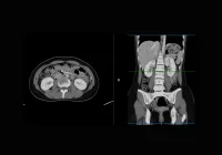
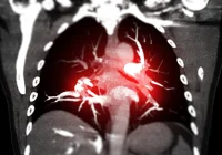
Segmentation in medical imaging has evolved into a critical tool for extracting accurate, quantitative insights from radiological data. With the increasing integration of artificial intelligence into healthcare, precise segmentation supports diagnosis, treatment planning and monitoring, and enables more reliable use of volumetric and radiomics analyses. Despite its importance, many radiologists lack practical guidance on how to perform segmentation efficiently and consistently across clinical and research settings. Recent recommendations from the European Society of Medical Imaging Informatics (EuSoMII) provide a structured framework to support radiologists and allied professionals in adopting best practices for segmentation. These recommendations cover technical standards, workflow optimisation and quality assurance, aiming to streamline segmentation procedures and improve consistency in outputs.
Establishing Robust Segmentation Protocols
A well-defined protocol forms the foundation of effective segmentation. It ensures the process aligns with clinical questions and research goals, standardises data input and reduces variability. Protocol development should be collaborative, involving radiologists and technical specialists to refine imaging parameters, software tools and evaluation metrics. Core components include patient selection criteria, imaging sequence preferences and documentation standards. Additionally, incorporating training resources such as visual atlases and checklists can enhance inter-reader consistency. Standardisation initiatives like CLAIM and CLEAR help ensure segmentation practices are transparent, reproducible and scientifically robust. In clinical contexts, standardised protocols are shown to reduce inter-reader variability, particularly in complex tasks such as target volume delineation for radiotherapy.
Must Read: Advancing Multiorgan Segmentation for Abdominal MRI
Maintaining segmentation consistency also requires preselecting specific imaging planes, sequences and contrast phases. Axial planes are generally preferred for their resolution, but alternative planes may be necessary depending on modality or lesion location. It is critical to choose the most appropriate MR sequence or CT phase before segmentation and to ensure consistency across datasets. Where multiple sequences are involved, radiologists must account for misalignments despite image registration efforts. Limiting the number of sequences per case, while retaining access to relevant reference sequences, balances workload with diagnostic accuracy.
Optimising Technical Parameters for Accurate Segmentation
High image quality underpins reliable segmentation. Suboptimal scans—due to low signal-to-noise ratios, artefacts or inappropriate reconstructions—can compromise segmentation performance. Radiologists should use reconstructions suited to the task: for example, soft tissue kernels in CT are better suited to lesion segmentation than sharp kernels, which introduce noise. Quantitative metrics like contrast-to-noise ratio and structural similarity index can complement expert judgement to evaluate image suitability.
Slice thickness also significantly influences segmentation accuracy. Thinner slices offer greater anatomical detail, crucial for tasks such as radiomics or brain metastasis analysis. However, they come at the cost of longer segmentation times and increased noise. Thicker slices, though quicker to process, are susceptible to partial volume effects and less suitable for high-resolution tasks. A balance must be struck based on the structure of interest and clinical objective. For example, organ volumetry may tolerate slices up to 5 mm, whereas detailed tumour analysis often demands thinner cuts. Wherever possible, all segmentations in a study should maintain uniform slice thickness to avoid introducing artificial variability.
Window settings—especially in CT imaging—should be standardised across all readers and timepoints. The choice of window level and width can drastically affect lesion visibility and perceived size, particularly in cases involving lung nodules or soft tissue lesions. Using consistent windowing parameters ensures that subtle but clinically relevant features are not missed or misinterpreted.
Maintaining Consistency and Managing Automation Risks
As segmentation tasks scale across multi-reader projects or institutions, consistency becomes increasingly important. Segmenters must receive adequate training, and workflows should include peer review and consensus discussions. Software tools and file formats should be standardised, ideally favouring DICOM SEG for voxel-based segmentations. Radiologists should be mindful of technical errors such as floating pixels or partial contours, which have minimal impact on volume estimation but can skew radiomics results.
AI and automation are increasingly used to accelerate segmentation, but they present specific risks. AI-assisted segmentation requires human oversight to correct errors and mitigate automation bias—the tendency to trust machine output unquestioningly. Overreliance on AI can lead to inaccuracies, especially among less experienced radiologists or in high-pressure environments. Introducing quality control frameworks that track both model performance and radiologist editing times helps detect such biases. If adjustment times drop while apparent accuracy rises, it may indicate overdependence on AI outputs rather than genuine model improvement.
High-quality initial segmentations are essential when training AI models. Accurate annotations improve model performance over time through iterative refinement. Radiologists can enhance both model training and research validity by ensuring clean, precise input data. Tools like TotalSegmentator offer reliable base segmentations that reduce radiologist workload, though human correction remains necessary to maintain quality.
Segmentation plays a vital role in the future of radiology, particularly as volumetric imaging and AI applications become more prominent. For radiologists, mastering this skill goes beyond technical execution; it requires a deep understanding of workflow design, imaging parameters and the implications of automation. Adopting standardised protocols, ensuring consistency in image handling and fostering collaboration between clinical and technical teams are essential to deliver accurate and reproducible results. Through practical recommendations and adherence to best practices, segmentation can become a more efficient, reliable tool for both clinical care and research, enhancing the precision and value of radiological work.
Source: European Radiology
Image Credit: iStock


