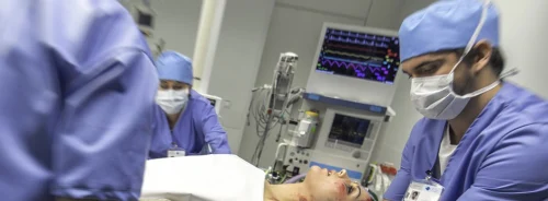
ICU Management & Practice, Volume 21 - Issue 5, 2021
The microbiota is recognised as one of the most important factors that can worsen the clinical conditions of patients who are already very frail in the intensive care unit. It also plays a crucial role in the prevention of ICU associated complications. It’s important to ensure the best functioning of the intestinal immune system.
The gastrointestinal tract represents one of the barriers between the external environment and the human body. This organ is composed of a wide variety of cells devoted to keep the intestine in balance (McDermott and Huffnagle 2014; Abreu 2010; del Rio et al. 2010; Sun et al. 2007).
The body's microbial composition directly influences the maturation of the immune response by GALT- Gut-Associated Lymphoid Tissue (Abreu 2010; del Rio 2010; Sun et al. 2007); and its continued effectiveness, protects against pathogen overgrowth, and modulates the balance between inflammation and immune homeostasis (Lynch and Pedersen 2016; Zhang and Frenette 2019). A few hours after admission to hospital, and especially to the intensive care unit (ICU), the intestinal microbiome switches to pathobiota (Stetcher et al. 2012; Babrowski et al. 2013; Hayakawa et al. 2011).
Sepsis can have a considerable impact on gastrointestinal function, indeed altered permeability and subsidence of normal intestinal flora can lead to systemic infection (Dickson 2015) such as neuropsychiatric disorders, inflammatory bowel disease (IBD), functional gastrointestinal (GI) disorders, cardiovascular disease, liver disease (Longhitano et al. 2020; Nakov et al. 2020) but also multiorgan failure (MOF) and severe sepsis.
The physiological mechanisms altered during hospitalisation in the ICU are responsible for:
- the growth of the pathobiote: colonisation by virulent bacteria;
- hypoperfusion of the intestinal tract leading to the massive release of nitrates and free oxygen radicals;
- the slow transit of faecal material due to various drugs (e.g. antibiotics, intravenous sedatives, opioids, catecholamines) causing a decrease in the normal turnover of about a trillion bacteria (Vincent et al. 2009);
- drugs such as proton pump inhibitors that change the chemical and physical characteristics of intestinal mucous, which neutralises gastric pH and reduces the velocity of gastric emptying (Marshall et al. 1993).
All this leads to the intestine colonisation by the Proteobacteria phylum (the most important are Pseudomonas aeruginosa and Escherischia coli and other minor gram-negative bacteria); also, other bacteria belonging to Firmicutes phylum (Staphylococcus aureus, Enterococcus spp and gram-positive bacteria such as Clostridium difficile) can colonise the intestine (Gootjans et al. 2010). The presence of the pathological bacteria described above (Marshall et al. 1993) is predictive of the development of MOF and systemic infections. Recently, the contribution of the microbiome in the development of coronary artery disease (CAD) has also been reported (Piccioni et al. 2021).
In addition to the above mentioned predisposing factors, the occurrence of contamination of the pulmonary ecosystem is also observed in intensive care patients as a result of the decrease in tussigenic stimulus, the migration of bacteria due to contiguity (Sands et al. 2017) through the orotracheal tube and the effect of microaspiration of bacteria from the oral cavity (Dickson et al. 2015; Gleeson et al. 1997; Segal et al. 2013; Sekizawa et al. 1990). All this leads to a subversion of the normal oral flora and leads to its replacement by pathogenic bacteria, such as Pseudomonas aeruginosa and Klebsiella pneumoniae.
It is obvious that one of the principal therapeutic goals in the ICU is to counterbalance dysbiosis. A number of treatment plans have been proposed: the use of probiotics in patients admitted to the ICU (Hill et al. 2014), faecal microbiota transplantation (FMT) (Fischer et al. 2017; Ianiro et al. 2017) and combination therapies that can help preserve mucosal integrity.
The use of probiotics as co-adjuvants, especially in intensive care patients, is rising exponentially thanks to studies conducted over the last 20 years, which show a statistically significant reduction in infections, in the incidence of VAP and, not least at all, in overall mortality.
A very recent 2020 review by Lukovic et al. (2019) highlighted some strategies that have been applied to reduce the risk of developing ventilator-associated pneumonia (VAP) such as oral decontamination, ETT impregnated with antimicrobials or silver, and probiotics (Spreadborough et al. 2016; Kollef et al. 2008; Muscedere et al. 2011; Manzaneres et al. 2016).
Also symbiotics (e.g. bifidobacterium breve strain yakult, lactobacillus casei strain Shirota, and galacto-oligosaccharides) are used in intensive care patients to prevent the development of sepsis. Conceptually they are a combination of prebiotics and probiotics and can be considered as enhancing compounds between the two and could be the best initial treatment during the conditions of altered homeostasis of the intestinal flora (Shimizu et al. 2018).
A new strategy in the treatment of chronic diarrhoea due to Clostridium difficile infection is FMT which has shown very strong results (Cammarota et al. 2017).
The microbiota is recognised as one of the most important factors that can worsen the clinical conditions of patients who are already very frail in the intensive care unit. At the same time, the microbiota also plays a crucial role in the prevention of ICU associated complications. It’s very important to use little but solid knowledge we have on the microbiome to ensure the best functioning of the intestinal immune system.
Also Rello et al. (2021) explained in their editorial the relation between pneumonia and dysbiosis that the traditional paradigm of VAP (that it is a disease caused by a single bacterial pathogen acquired through microaspiration) needs to be replaced by a hypothetical model in which VAP would be associated with dysbiosis. The gut microbiome contributes to protect against opportunistic pathogens. Enriching the microbiota with members of the phylum Proteobacteria, which are considered commensals, increases serum IgA levels. Thus, lung dysbiosis combined with gut dysbiosis might induce local immunosuppression and lung dysfunction, facilitating the occurrence of VAP. The role of the Th17 response provoked by segmented filamentous bacteria, which provides protection from staphylococcal pneumonia, seems crucial. These observations are not only of academic interest. Early identification of patients with dysbiosis associated with a higher risk of developing VAP is an unmet clinical need, and this should lead to innovative, targeted preventive strategies (Cammarota et al. 2017).
Conflict of Interest
None.
References:
Abreu MT (2010) Toll-like receptor signalling in the intestinal epithelium: how bacterial recognition shapes intestinal function. Nat Rev Immunol, 10:131-44.
Babrowski T, Romanowski K, Fink D et al. (2013) The intestinal environment of surgical injury transforms Pseudomonas aeruginosa into a discrete hypervirulent morphotype capable of causing lethal peritonitis. Surgery, 153(1):36–43.
Cammarota G, Ianiro G, Tilg H et al. (2017) European consensus conference on faecal microbiota transplantation in clinical practice. Gut, 66(4):569-580.
del Rio ML, Bernhardt G, Rodriguez-Barbosa JI, Forster R (2010) Development and functional specialization of CD103+ dendritic cells. Immunol Rev, 234:268–81
Dickson RP (2016) The microbiome and critical illness. Lancet Respir Med, 4(1):59-72.
Dickson RP, Erb-Downward JR, Freeman CM et al. (2015) Spatial Variation in the healthy human lung microbiome and the adapted island model of lung biogeography. Ann Am Thorac Soc, 12:821–30.
Fischer M, Sipe B, ChengY-W et al. (2017) Fecal microbiota transplant in severe and severe-complicated Clostridium difficile: a promising treatment approach. Gut Microbes, 8:289e302.
Gleeson K, Eggli DF, Maxwell SL (1997) Quantitative aspiration during sleep in normal subjects. Chest, 111:1266–72.
Gootjans J, Lenaerts K,Derikx JP et al. (2010) , Human intestinal ischemia-reperfusion- induced inflammation characterized: experiences from a new translational model. Am J Pathol, 176:2283–91.
Hayakawa M, Asahara T, Henzan N et al. (2011) Dramatic changes of the gut flora immediately after severe and sudden insults. Dig Dis Sci, 56(8):2361–5.
Hill C, Guarner F, Reid G et al. (2014) Gibson GR, Merenstein DJ, Pot B, Morelli L, Canani RB, Flint HJ, Salminen S, Calder PC, Sanders ME. Expert consensus document. The International Scientific Association for Probiotics and Prebiotics consensus statement on the scope and appropriate use of the term probiotic. Nat Rev Gastroenterol Hepatol, 11(8):506-14.
Ianiro G, Valerio L, Masucci L et al. (2017) Predictors of failure after single faecal microbiota transplantation in patients with recur- rent Clostridium difficile infection: results from a 3-year, single-centre cohort study. Clin Microbiol Infect, 23:337.e1e3.
Kollef MH, Afessa B, Anzueto A et al. (2008) Silver-coated endotracheal tubes and incidence of ventilator-associated pneumonia: the NASCENT randomized trial. JAMA, 300:805-813.
Longhitano Y, Zanza C, Thangathurai D et al. (2020 Gut Alterations in Septic Patients: A Biochemical Literature Review. Rev Recent Clin Trials, 15(4):289-297.
Lukovic E, Moitra VK, Freedberg DE (2019) The microbiome: implications for perioperative and critical care. Curr Opin Anaesthesiol, 32(3):412-420.
Lynch SV, Pedersen O (2016) The Human Intestinal Microbiome in Health and Disease. N Engl J Med, 15;375(24):2369-2379.
Manzanares W, Lemieux M, Langlois PI et al. (2016) Probiotic and synbiotic therapy in critical illness: a systematic review and meta-analysis. Crit Care, 19:262.
Marshall JC, Christou NV, Meakins JL (1993) The gastrointestinal tract. The “undrained abscess” of multiple organ failure. Ann Surg, 218:111–9.
McDermott AJ, Huffnagle GB (2014) The microbiome and regulation of mucosal immunity. Immunology, 142(1):24-31.
Muscedere J, Rewa O, McKechnie K et al. (2011) Subglottic secretion drainage for the prevention of ventilator-associated pneumonia: a systematic review and meta- analysis. Crit Care Med, 39:1985–91.
Nakov R, Segal JP, Settanni CR et al. (2020) Microbiome: what intensivists should know. Minerva Anestesiol, 86(7):777-785.
Piccioni A, de Cunzo T, Valletta F et al. (2021) Gut microbiota and environment in coronary artery disease. Int. J. Environ. Res. Public Health, 18.
Rello J, Schrenzel J, Tejo AM (2021) New insights into pneumonia in patients on prolonged mechanical ventilation: need for a new paradigm addressing dysbiosis. J Bras Pneumol, 47(3):e20210198.
Sands KM, Wilson MJ, Lewis MAO et al. (2017) Respiratory pathogen colonization of dental plaque, the lower airways, and endotracheal tube biofilms during mechanical ventilation. J Crit Care, 37:30 –37.
Segal LN, Alekseyenko AV, Clemente JC et al. (2013) Enrichment of lung microbiome with supraglottic taxa is associated with increased pulmonary inflammation. Microbiome, 1:19.
Sekizawa K, Ujiie Y, Itabashi S et al. (1990) Sasaki H, Takishima T. Lack of cough reflex in aspiration pneumonia. Lancet, 335:1228–29.
Spreadborough P, Lort S, Pasquali S et al. (2016) A systematic review and meta- analysis of perioperative oral decontamination in patients undergoing major elective surgery. Perioper Med (Lond), 5:6.
Shimizu K, Yamada T, Ogura H et al. (2018) Synbiotics modulate gut microbiota and reduce enteritis and ventilator-associated pneumonia in patients with sepsis: a randomized controlled trial. Crit Care, 22(1):239.
Stecher B, Denzler R, Maier L et al. (2012) Gut inflammation can boost horizontal gene transfer between pathogenic and commensal Enterobacteriaceae. Proc Natl Acad Sci USA., 109(4):1269–74.
Sun CM, Hall JA, Blank RB et al. (2007) Small intestine lamina propria dendritic cells promote de novo generation of Foxp3 Treg cells via retinoic acid. J Exp Med 2007; 204:1775–85.
Vincent JL, Rello J, Marshall J et al. (2009) EPIC II Group of Investigators. International study of the prevalence and outcomes of infection in intensive care units. JAMA, 302:2323–2329.
Zhang D, Frenette PS (2019) Cross talk between neutrophils and the microbiota. Blood, 133(20):2168-2177.


