A new type of medical imaging technology that could diagnose cardiovascular disease by measuring ultrasound signals from molecules exposed to a fast-pulsing laser is now being commercialised. According to researchers, the system takes precise three-dimensional images of plaques lining arteries and identifies deposits that may rupture and cause heart attacks. Their findings are published online in the Nature journal Scientific Reports.
The new imaging technique reveals the presence of carbon-hydrogen bonds making up lipid molecules in arterial plaques that cause heart disease. "This allows us to see the exact nature of plaque formation in the walls of arteries so we can define whether plaque is going to rupture," said Michael Sturek, co-author of the study and a professor and chair of the Department of Cellular & Integrative Physiology at Indiana University School of Medicine.
Laser-Based Intravascular Photoacoustic Imaging
Some plaques are more dangerous than others, and it is important to know the chemical makeup of the blood vessel wall "to determine which ones are at risk of rupturing," Sturek noted. However, research in the area has been hampered by the inability to perform high-speed imaging in tissue. To overcome the problem, the researchers developed a Raman laser using a laser that produces 2,000 pulses per second. Each pulse is able to generate an image, representing a 100-fold increase in the imaging speed of the new technology, called intravascular photoacoustic imaging.
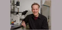
"This innovation represents a big step toward advancing this technology to the clinical setting," said Ji-Xin Cheng, a professor in Purdue University's Weldon School of Biomedical Engineering and Department of Chemistry. The collaborative work included researchers from Purdue, Indiana University School of Medicine, the University of Southern California, the University of California at Irvine, and startup company Spectral Energy.
The laser, which pulses in the near-infrared range of the spectrum, causes tissue to heat and expand locally, generating pressure waves at the ultrasound frequency that can be picked up with a device called a transducer, the research team explained. The near-infrared laser causes enough heating to generate ultrasound but not enough to damage the tissues. The research was conducted with intact pig tissue and will expand to research with live animals and then clinical studies with humans.
‘Label Free’ Imaging Technique is Ideal for Diagnostic Uses
The "label free" imaging technique does not require samples to be marked with dyes, making it ideal for diagnostic applications. Also, the system is small enough to be incorporated into an endoscope to put into blood vessels using a catheter, Cheng said.
The new imaging technology is being commercialised by the company Vibronix Inc., co-founded by Cheng and Purdue postdoctoral research associate Pu Wang. The Purdue Office of Technology Commercialization has filed a US patent application.
The study was authored by Wang; Purdue research scientist Mikhail N. Slipchenko; Purdue graduate student Jie Hui; USC graduate students Teng Ma and Shanshan Liang; USC researcher Qifa Zhou; K. Kirk Shung, director of the Ultrasonic Transducer Resource Center at USC; Sukesh Roy, CEO of Spectral Energy LLC in Dayton, Ohio; Sturek; Zhongping Chen, a researcher at UC Irvine; and Cheng.
Funding sources for the research included the National Institutes of Health and the American Heart Association.
Source: ScienceDaily.com
Image Credit: Purdue University Weldon School of Biomedical Engineering/Ji-Xin Cheng
The new imaging technique reveals the presence of carbon-hydrogen bonds making up lipid molecules in arterial plaques that cause heart disease. "This allows us to see the exact nature of plaque formation in the walls of arteries so we can define whether plaque is going to rupture," said Michael Sturek, co-author of the study and a professor and chair of the Department of Cellular & Integrative Physiology at Indiana University School of Medicine.
Laser-Based Intravascular Photoacoustic Imaging
Some plaques are more dangerous than others, and it is important to know the chemical makeup of the blood vessel wall "to determine which ones are at risk of rupturing," Sturek noted. However, research in the area has been hampered by the inability to perform high-speed imaging in tissue. To overcome the problem, the researchers developed a Raman laser using a laser that produces 2,000 pulses per second. Each pulse is able to generate an image, representing a 100-fold increase in the imaging speed of the new technology, called intravascular photoacoustic imaging.
"This innovation represents a big step toward advancing this technology to the clinical setting," said Ji-Xin Cheng, a professor in Purdue University's Weldon School of Biomedical Engineering and Department of Chemistry. The collaborative work included researchers from Purdue, Indiana University School of Medicine, the University of Southern California, the University of California at Irvine, and startup company Spectral Energy.
The laser, which pulses in the near-infrared range of the spectrum, causes tissue to heat and expand locally, generating pressure waves at the ultrasound frequency that can be picked up with a device called a transducer, the research team explained. The near-infrared laser causes enough heating to generate ultrasound but not enough to damage the tissues. The research was conducted with intact pig tissue and will expand to research with live animals and then clinical studies with humans.
‘Label Free’ Imaging Technique is Ideal for Diagnostic Uses
The "label free" imaging technique does not require samples to be marked with dyes, making it ideal for diagnostic applications. Also, the system is small enough to be incorporated into an endoscope to put into blood vessels using a catheter, Cheng said.
The new imaging technology is being commercialised by the company Vibronix Inc., co-founded by Cheng and Purdue postdoctoral research associate Pu Wang. The Purdue Office of Technology Commercialization has filed a US patent application.
The study was authored by Wang; Purdue research scientist Mikhail N. Slipchenko; Purdue graduate student Jie Hui; USC graduate students Teng Ma and Shanshan Liang; USC researcher Qifa Zhou; K. Kirk Shung, director of the Ultrasonic Transducer Resource Center at USC; Sukesh Roy, CEO of Spectral Energy LLC in Dayton, Ohio; Sturek; Zhongping Chen, a researcher at UC Irvine; and Cheng.
Funding sources for the research included the National Institutes of Health and the American Heart Association.
Source: ScienceDaily.com
Image Credit: Purdue University Weldon School of Biomedical Engineering/Ji-Xin Cheng
Latest Articles
Ultrasound, heart attack, arterial plaque, cardiovascular disease, photoacoustics
A new type of medical imaging technology that could diagnose cardiovascular disease by measuring ultrasound signals from molecules exposed to a fast-pulsin...